6QB9 | pdb_00006qb9

Structure of an anti-Mcl1 scFv


Experimental Data Snapshot

  • Method: X-RAY DIFFRACTION
  • Resolution: 1.85 Å
  • R-Value Free: 
    0.239 (Depositor), 0.250 (DCC) 
  • R-Value Work: 
    0.200 (Depositor), 0.203 (DCC) 
  • R-Value Observed: 
    0.202 (Depositor) 

wwPDB Validation 3D Report Full Report

Validation slider image for 6QB9

Ligand Structure Quality Assessment 


This is version 1.2 of the entry. See complete history

Literature

Antibody fragments structurally enable a drug-discovery campaign on the cancer target Mcl-1.

Luptak, J.Bista, M.Fisher, D.Flavell, L.Gao, N.Wickson, K.Kazmirski, S.L.Howard, T.Rawlins, P.B.Hargreaves, D.

(2019) Acta Crystallogr D Struct Biol 75: 1003-1014

  • DOI: https://doi.org/10.1107/S2059798319014116
  • Primary Citation Related Structures: 
    6QB3, 6QB4, 6QB6, 6QB9, 6QBC, 6QF9, 6QFC

  • PubMed Abstract: 

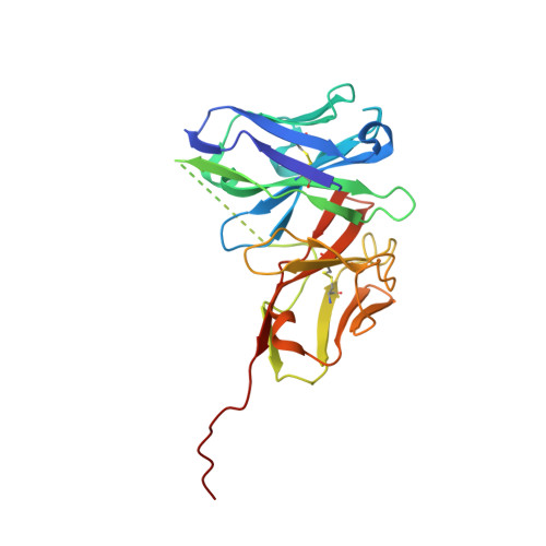
    Apoptosis is a crucial process by which multicellular organisms control tissue growth, removal and inflammation. Disruption of the normal apoptotic function is often observed in cancer, where cell death is avoided by the overexpression of anti-apoptotic proteins of the Bcl-2 (B-cell lymphoma 2) family, including Mcl-1 (myeloid cell leukaemia 1). This makes Mcl-1 a potential target for drug therapy, through which normal apoptosis may be restored by inhibiting the protective function of Mcl-1. Here, the discovery and biophysical properties of an anti-Mcl-1 antibody fragment are described and the utility of both the scFv and Fab are demonstrated in generating an Mcl-1 crystal system amenable to iterative structure-guided drug design.


  • Organizational Affiliation
    • Discovery Sciences, R&D Biopharmaceuticals, AstraZeneca, Cambridge CB4 0WG, England.

Macromolecule Content 

  • Total Structure Weight: 53.16 kDa 
  • Atom Count: 3,758 
  • Modeled Residue Count: 473 
  • Deposited Residue Count: 510 
  • Unique protein chains: 1

Macromolecules

Find similar proteins by:|  3D Structure
Entity ID: 1
MoleculeChains  Sequence LengthOrganismDetailsImage
scFv55A,
B [auth X]
255Homo sapiensMutation(s): 0 

Experimental Data & Validation

Experimental Data

  • Method: X-RAY DIFFRACTION
  • Resolution: 1.85 Å
  • R-Value Free:  0.239 (Depositor), 0.250 (DCC) 
  • R-Value Work:  0.200 (Depositor), 0.203 (DCC) 
  • R-Value Observed: 0.202 (Depositor) 
Space Group: C 1 2 1
Unit Cell:
Length ( Å )Angle ( ˚ )
a = 77.88α = 90
b = 63.03β = 109.96
c = 101.7γ = 90
Software Package:
Software NamePurpose
BUSTERrefinement
MOSFLMdata reduction
Aimlessdata scaling
PHASERphasing

Structure Validation

View Full Validation Report


Ligand Structure Quality Assessment 


Entry History 

Deposition Data

Revision History  (Full details and data files)

  • Version 1.0: 2019-11-06
    Type: Initial release
  • Version 1.1: 2019-11-13
    Changes: Database references
  • Version 1.2: 2024-11-20
    Changes: Data collection, Database references, Structure summary