6Q9E | pdb_00006q9e

Complex III2 focused refinement from Ovine respiratory supercomplex I+III2


Experimental Data Snapshot

  • Method: ELECTRON MICROSCOPY
  • Resolution: 3.90 Å
  • Aggregation State: PARTICLE 
  • Reconstruction Method: SINGLE PARTICLE 

Starting Model: experimental
View more details

wwPDB Validation 3D Report Full Report

Validation slider image for 6Q9E

This is version 1.3 of the entry. See complete history

Literature

Structures of Respiratory Supercomplex I+III2Reveal Functional and Conformational Crosstalk.

Letts, J.A.Fiedorczuk, K.Degliesposti, G.Skehel, M.Sazanov, L.A.

(2019) Mol Cell 75: 1131-1146.e6

  • DOI: https://doi.org/10.1016/j.molcel.2019.07.022
  • Primary Citation Related Structures: 
    6Q9B, 6Q9D, 6Q9E, 6QA9, 6QBX, 6QC2, 6QC3, 6QC4, 6QC5, 6QC6, 6QC7, 6QC8, 6QC9, 6QCA, 6QCF

  • PubMed Abstract: 

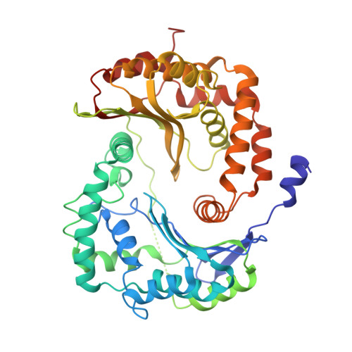
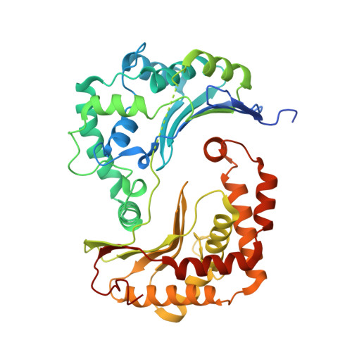
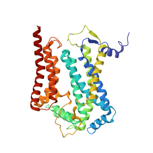
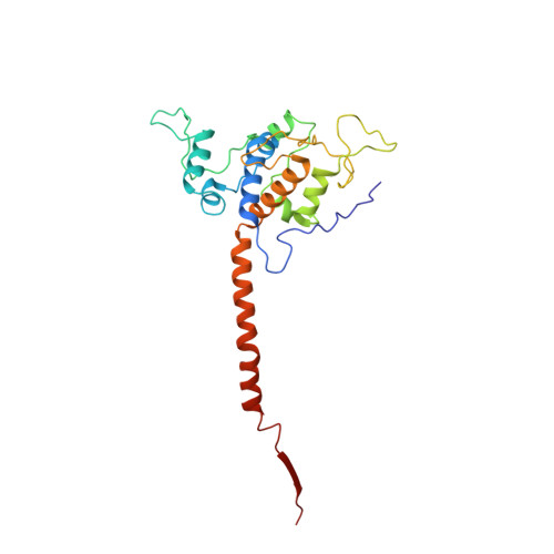
    The mitochondrial electron transport chain complexes are organized into supercomplexes (SCs) of defined stoichiometry, which have been proposed to regulate electron flux via substrate channeling. We demonstrate that CoQ trapping in the isolated SC I+III 2 limits complex (C)I turnover, arguing against channeling. The SC structure, resolved at up to 3.8 Å in four distinct states, suggests that CoQ oxidation may be rate limiting because of unequal access of CoQ to the active sites of CIII 2 . CI shows a transition between "closed" and "open" conformations, accompanied by the striking rotation of a key transmembrane helix. Furthermore, the state of CI affects the conformational flexibility within CIII 2 , demonstrating crosstalk between the enzymes. CoQ was identified at only three of the four binding sites in CIII 2 , suggesting that interaction with CI disrupts CIII 2 symmetry in a functionally relevant manner. Together, these observations indicate a more nuanced functional role for the SCs.


  • Organizational Affiliation
    • Institute of Science and Technology Austria, Klosterneuberg 3400, Austria; Department of Molecular and Cellular Biology, University of California, Davis, Davis, CA 95616, USA.

Macromolecule Content 

  • Total Structure Weight: 477.55 kDa 
  • Atom Count: 31,997 
  • Modeled Residue Count: 3,988 
  • Deposited Residue Count: 4,130 
  • Unique protein chains: 10

Macromolecules

Find similar proteins by:|  3D Structure
Entity ID: 1
MoleculeChains  Sequence LengthOrganismDetailsImage
Ubiquinol-cytochrome c reductase core protein 1A [auth a1],
K [auth a3]
446Ovis ariesMutation(s): 0 
Find similar proteins by:|  3D Structure
Entity ID: 2
MoleculeChains  Sequence LengthOrganismDetailsImage
Ubiquinol-cytochrome c reductase core protein 2B [auth a2],
L [auth a4]
439Ovis ariesMutation(s): 0 
Find similar proteins by:|  3D Structure
Entity ID: 3
MoleculeChains  Sequence LengthOrganismDetailsImage
Cytochrome bC [auth b1],
M [auth b2]
379Ovis ariesMutation(s): 0 
Membrane Entity: Yes 
UniProt
Find proteins for P24959 (Ovis aries)
Explore P24959 
Go to UniProtKB:  P24959
Entity Groups
Sequence Clusters30% Identity50% Identity70% Identity90% Identity95% Identity100% Identity
UniProt GroupP24959
Sequence Annotations
Expand
Reference Sequence
Find similar proteins by:|  3D Structure
Entity ID: 4
MoleculeChains  Sequence LengthOrganismDetailsImage
Cytochrome c1D [auth c1],
N [auth c2]
240Ovis ariesMutation(s): 0 
Find similar proteins by:|  3D Structure
Entity ID: 5
MoleculeChains  Sequence LengthOrganismDetailsImage
Cytochrome b-c1 complex subunit Rieske, mitochondrialE [auth f1],
O [auth f2]
196Ovis ariesMutation(s): 0 
EC: 1.10.2.2
Find similar proteins by:|  3D Structure
Entity ID: 6
MoleculeChains  Sequence LengthOrganismDetailsImage
Cytochrome b-c1 complex subunit 7F [auth d1],
P [auth d2]
110Ovis ariesMutation(s): 0 
Find similar proteins by:|  3D Structure
Entity ID: 7
MoleculeChains  Sequence LengthOrganismDetailsImage
Ubiquinol-cytochrome c reductase complex III subunit VIIG [auth q1],
Q [auth q2]
81Ovis ariesMutation(s): 0 
Find similar proteins by:|  3D Structure
Entity ID: 8
MoleculeChains  Sequence LengthOrganismDetailsImage
Cytochrome b-c1 complex subunit 6H [auth h1],
R [auth h2]
78Ovis ariesMutation(s): 0 
Find similar proteins by:|  3D Structure
Entity ID: 9
MoleculeChains  Sequence LengthOrganismDetailsImage
Cytochrome b-c1 complex subunit Rieske, mitochondrialI [auth x1],
S [auth x2]
33Ovis ariesMutation(s): 0 
Find similar proteins by:|  3D Structure
Entity ID: 10
MoleculeChains  Sequence LengthOrganismDetailsImage
Ubiquinol-cytochrome c reductase, complex III subunit XJ [auth i1],
T [auth i2]
63Ovis ariesMutation(s): 0 

Small Molecules

Ligands 6 Unique
IDChains Name / Formula / InChI Key2D Diagram3D Interactions
CDL

Query on CDL


Download:Ideal Coordinates CCD File
DA [auth c1],
IA [auth b2],
LA [auth c2],
X [auth b1],
Y [auth b1]
CARDIOLIPIN
C81 H156 O17 P2
XVTUQDWPJJBEHJ-KZCWQMDCSA-L
U10

Query on U10


Download:Ideal Coordinates CCD File
AA [auth b1],
JA [auth b2],
Z [auth b1]
UBIQUINONE-10
C59 H90 O4
ACTIUHUUMQJHFO-UPTCCGCDSA-N
3PE

Query on 3PE


Download:Ideal Coordinates CCD File
CA [auth c1],
HA [auth b2],
MA [auth f2],
W [auth b1]
1,2-Distearoyl-sn-glycerophosphoethanolamine
C41 H82 N O8 P
LVNGJLRDBYCPGB-LDLOPFEMSA-N
HEC

Query on HEC


Download:Ideal Coordinates CCD File
BA [auth c1],
KA [auth c2]
HEME C
C34 H34 Fe N4 O4
HXQIYSLZKNYNMH-LJNAALQVSA-N
HEM

Query on HEM


Download:Ideal Coordinates CCD File
FA [auth b2],
GA [auth b2],
U [auth b1],
V [auth b1]
PROTOPORPHYRIN IX CONTAINING FE
C34 H32 Fe N4 O4
KABFMIBPWCXCRK-RGGAHWMASA-L
FES

Query on FES


Download:Ideal Coordinates CCD File
EA [auth f1],
NA [auth f2]
FE2/S2 (INORGANIC) CLUSTER
Fe2 S2
NIXDOXVAJZFRNF-UHFFFAOYSA-N

Experimental Data & Validation

Experimental Data

  • Method: ELECTRON MICROSCOPY
  • Resolution: 3.90 Å
  • Aggregation State: PARTICLE 
  • Reconstruction Method: SINGLE PARTICLE 
EM Software:
TaskSoftware PackageVersion
RECONSTRUCTIONRELION2
MODEL REFINEMENTPHENIX1.13-2998

Structure Validation

View Full Validation Report


Entry History 

& Funding Information

Deposition Data


Funding OrganizationLocationGrant Number
European Research CouncilAustria701309

Revision History  (Full details and data files)

  • Version 1.0: 2019-08-21
    Type: Initial release
  • Version 1.1: 2019-09-18
    Changes: Data collection, Database references
  • Version 1.2: 2019-10-02
    Changes: Data collection, Database references
  • Version 1.3: 2024-11-13
    Changes: Data collection, Database references, Derived calculations, Refinement description, Structure summary