6Q5E | pdb_00006q5e

Crystal structure of the ferric enterobactin receptor from Pseudomonas aeruginosa (PfeA) in complex with enterobactin


Experimental Data Snapshot

  • Method: X-RAY DIFFRACTION
  • Resolution: 2.70 Å
  • R-Value Free: 
    0.249 (Depositor), 0.249 (DCC) 
  • R-Value Work: 
    0.208 (Depositor), 0.210 (DCC) 
  • R-Value Observed: 
    0.210 (Depositor) 

Starting Model: experimental
View more details

wwPDB Validation 3D Report Full Report

Validation slider image for 6Q5E

Ligand Structure Quality Assessment 


This is version 1.3 of the entry. See complete history

Literature

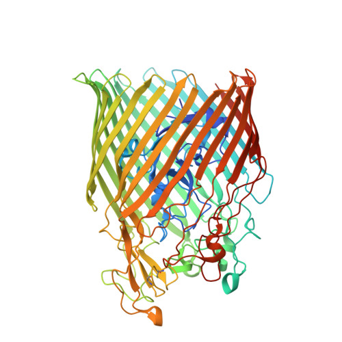
The complex of ferric-enterobactin with its transporter from Pseudomonas aeruginosa suggests a two-site model.

Moynie, L.Milenkovic, S.Mislin, G.L.A.Gasser, V.Malloci, G.Baco, E.McCaughan, R.P.Page, M.G.P.Schalk, I.J.Ceccarelli, M.Naismith, J.H.

(2019) Nat Commun 10: 3673-3673

  • DOI: https://doi.org/10.1038/s41467-019-11508-y
  • Primary Citation Related Structures: 
    5M9B, 5MZS, 5NC4, 5NR2, 5OUT, 6I2J, 6Q5E, 6R1F

  • PubMed Abstract: 

    Bacteria use small molecules called siderophores to scavenge iron. Siderophore-Fe 3+ complexes are recognised by outer-membrane transporters and imported into the periplasm in a process dependent on the inner-membrane protein TonB. The siderophore enterobactin is secreted by members of the family Enterobacteriaceae, but many other bacteria including Pseudomonas species can use it. Here, we show that the Pseudomonas transporter PfeA recognises enterobactin using extracellular loops distant from the pore. The relevance of this site is supported by in vivo and in vitro analyses. We suggest there is a second binding site deeper inside the structure and propose that correlated changes in hydrogen bonds link binding-induced structural re-arrangements to the structural adjustment of the periplasmic TonB-binding motif.


  • Organizational Affiliation
    • Division of Structural Biology, Wellcome Trust Centre of Human Genomics, 7 Roosevelt Drive, Oxford, OX3 7BN, UK.

Macromolecule Content 

  • Total Structure Weight: 79.92 kDa 
  • Atom Count: 5,486 
  • Modeled Residue Count: 698 
  • Deposited Residue Count: 721 
  • Unique protein chains: 1

Macromolecules

Find similar proteins by:|  3D Structure
Entity ID: 1
MoleculeChains  Sequence LengthOrganismDetailsImage
Ferric enterobactin receptor721Pseudomonas aeruginosa PAO1Mutation(s): 0 
Gene Names: pfeAPA2688
Membrane Entity: Yes 
UniProt
Find proteins for Q05098 (Pseudomonas aeruginosa (strain ATCC 15692 / DSM 22644 / CIP 104116 / JCM 14847 / LMG 12228 / 1C / PRS 101 / PAO1))
Explore Q05098 
Go to UniProtKB:  Q05098
Entity Groups
Sequence Clusters30% Identity50% Identity70% Identity90% Identity95% Identity100% Identity
UniProt GroupQ05098
Sequence Annotations
Expand
Reference Sequence

Small Molecules

Ligands 3 Unique
IDChains Name / Formula / InChI Key2D Diagram3D Interactions
LP5

Query on LP5


Download:Ideal Coordinates CCD File
D [auth A](R)-((2R,3S,4R,5R,6R)-3-HYDROXY-2-(HYDROXYMETHYL)-5-((R)-3-HYDROXYTETRADECANAMIDO)-6-(PHOSPHONOOXY)TETRAHYDRO-2H-PYRAN-4-YL) 3-HYDROXYTETRADECANOATE
C34 H66 N O12 P
HEHQDWUWJVPREQ-XQJZMFRCSA-N
EB4

Query on EB4


Download:Ideal Coordinates CCD File
C [auth A]N,N',N''-[(3S,7S,11S)-2,6,10-trioxo-1,5,9-trioxacyclododecane-3,7,11-triyl]tris(2,3-dihydroxybenzamide)
C30 H27 N3 O15
SERBHKJMVBATSJ-BZSNNMDCSA-N
FE

Query on FE


Download:Ideal Coordinates CCD File
B [auth A]FE (III) ION
Fe
VTLYFUHAOXGGBS-UHFFFAOYSA-N

Experimental Data & Validation

Experimental Data

  • Method: X-RAY DIFFRACTION
  • Resolution: 2.70 Å
  • R-Value Free:  0.249 (Depositor), 0.249 (DCC) 
  • R-Value Work:  0.208 (Depositor), 0.210 (DCC) 
  • R-Value Observed: 0.210 (Depositor) 
Space Group: P 21 21 2
Unit Cell:
Length ( Å )Angle ( ˚ )
a = 85.63α = 90
b = 159.26β = 90
c = 78.61γ = 90
Software Package:
Software NamePurpose
REFMACrefinement
XDSdata reduction
XSCALEdata scaling
PHASERphasing

Structure Validation

View Full Validation Report


Ligand Structure Quality Assessment 


Entry History 

& Funding Information

Deposition Data


Funding OrganizationLocationGrant Number
United Kingdom--

Revision History  (Full details and data files)

  • Version 1.0: 2019-01-16
    Type: Initial release
  • Version 1.1: 2019-08-28
    Changes: Data collection, Database references
  • Version 1.2: 2024-01-24
    Changes: Data collection, Database references, Refinement description
  • Version 1.3: 2024-10-09
    Changes: Structure summary