6Q1Z | pdb_00006q1z

Crystal structure of human 1G04 Fab in complex with influenza virus neuraminidase from A/Hunan/02650/2016 (H7N9)


Experimental Data Snapshot

  • Method: X-RAY DIFFRACTION
  • Resolution: 3.45 Å
  • R-Value Free: 
    0.247 (Depositor), 0.257 (DCC) 
  • R-Value Work: 
    0.197 (Depositor), 0.210 (DCC) 
  • R-Value Observed: 
    0.200 (Depositor) 

Starting Models: experimental
View more details

wwPDB Validation 3D Report Full Report

Validation slider image for 6Q1Z

Ligand Structure Quality Assessment 


This is version 2.2 of the entry. See complete history

Literature

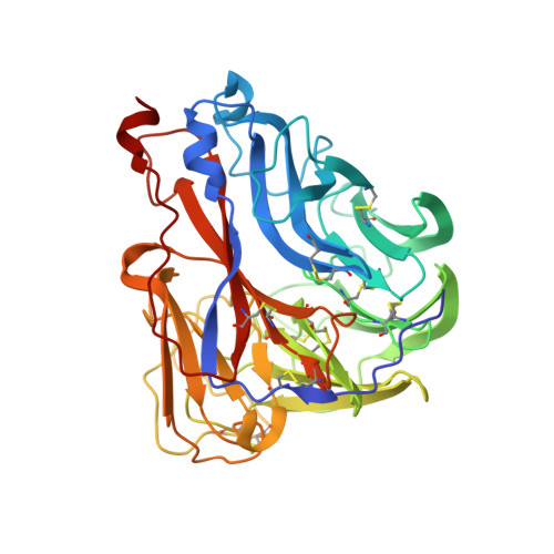
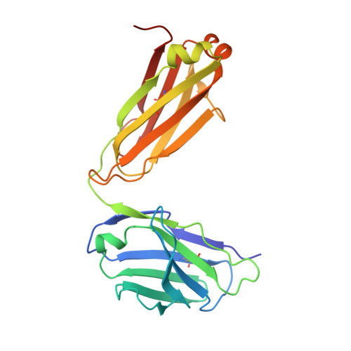
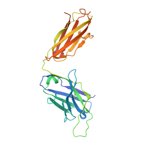
Broadly protective human antibodies that target the active site of influenza virus neuraminidase.

Stadlbauer, D.Zhu, X.McMahon, M.Turner, J.S.Wohlbold, T.J.Schmitz, A.J.Strohmeier, S.Yu, W.Nachbagauer, R.Mudd, P.A.Wilson, I.A.Ellebedy, A.H.Krammer, F.

(2019) Science 366: 499-504

  • DOI: https://doi.org/10.1126/science.aay0678
  • Primary Citation Related Structures: 
    6Q1Z, 6Q20, 6Q23

  • PubMed Abstract: 

    Better vaccines against influenza virus are urgently needed to provide broader protection against diverse strains, subtypes, and types. Such efforts are assisted by the identification of novel broadly neutralizing epitopes targeted by protective antibodies. Influenza vaccine development has largely focused on the hemagglutinin, but the other major surface antigen, the neuraminidase, has reemerged as a potential target for universal vaccines. We describe three human monoclonal antibodies isolated from an H3N2-infected donor that bind with exceptional breadth to multiple different influenza A and B virus neuraminidases. These antibodies neutralize the virus, mediate effector functions, are broadly protective in vivo, and inhibit neuraminidase activity by directly binding to the active site. Structural and functional characterization of these antibodies will inform the development of neuraminidase-based universal vaccines against influenza virus.


  • Organizational Affiliation
    • Department of Microbiology, Icahn School of Medicine at Mount Sinai, New York, NY 10029, USA.

Macromolecule Content 

  • Total Structure Weight: 192.78 kDa 
  • Atom Count: 13,236 
  • Modeled Residue Count: 1,660 
  • Deposited Residue Count: 1,698 
  • Unique protein chains: 3

Macromolecules

Find similar proteins by:|  3D Structure
Entity ID: 1
MoleculeChains  Sequence LengthOrganismDetailsImage
Neuraminidase
A, B
393Influenza A virusMutation(s): 0 
Gene Names: NA
EC: 3.2.1.18
UniProt
Find proteins for A0A5A4LHS4 (Influenza A virus)
Explore A0A5A4LHS4 
Go to UniProtKB:  A0A5A4LHS4
Entity Groups
Sequence Clusters30% Identity50% Identity70% Identity90% Identity95% Identity100% Identity
UniProt GroupA0A5A4LHS4
Glycosylation
Glycosylation Sites: 3
Sequence Annotations
Expand
Reference Sequence
Find similar proteins by:|  3D Structure
Entity ID: 2
MoleculeChains  Sequence LengthOrganismDetailsImage
1G04 Fab kappa light chainC [auth L],
E [auth C]
216Homo sapiensMutation(s): 0 
Find similar proteins by:|  3D Structure
Entity ID: 3
MoleculeChains  Sequence LengthOrganismDetailsImage
1G04 Fab IgG1 heavy chainD [auth H],
F [auth D]
240Homo sapiensMutation(s): 0 

Oligosaccharides

Help  
Entity ID: 4
MoleculeChains Length2D Diagram GlycosylationD Interactions
alpha-D-mannopyranose-(1-2)-alpha-D-mannopyranose-(1-2)-alpha-D-mannopyranose-(1-3)-[alpha-D-mannopyranose-(1-2)-alpha-D-mannopyranose-(1-3)-[alpha-D-mannopyranose-(1-2)-alpha-D-mannopyranose-(1-6)]alpha-D-mannopyranose-(1-6)]beta-D-mannopyranose-(1-4)-2-acetamido-2-deoxy-beta-D-glucopyranose-(1-4)-2-acetamido-2-deoxy-beta-D-glucopyranoseG [auth E],
I [auth G]
11N-Glycosylation
Glycosylation Resources
GlyTouCan: G60230HH
GlyCosmos: G60230HH
GlyGen: G60230HH
Entity ID: 5
MoleculeChains Length2D Diagram GlycosylationD Interactions
beta-D-mannopyranose-(1-4)-2-acetamido-2-deoxy-beta-D-glucopyranose-(1-4)-2-acetamido-2-deoxy-beta-D-glucopyranoseH [auth F],
J [auth I]
3N-Glycosylation
Glycosylation Resources
GlyTouCan: G15407YE
GlyCosmos: G15407YE
GlyGen: G15407YE

Experimental Data & Validation

Experimental Data

  • Method: X-RAY DIFFRACTION
  • Resolution: 3.45 Å
  • R-Value Free:  0.247 (Depositor), 0.257 (DCC) 
  • R-Value Work:  0.197 (Depositor), 0.210 (DCC) 
  • R-Value Observed: 0.200 (Depositor) 
Space Group: I 4
Unit Cell:
Length ( Å )Angle ( ˚ )
a = 180.242α = 90
b = 180.242β = 90
c = 148.125γ = 90
Software Package:
Software NamePurpose
PHENIXrefinement
HKL-2000data reduction
HKL-2000data scaling
PHASERphasing

Structure Validation

View Full Validation Report


Ligand Structure Quality Assessment 


Entry History 

& Funding Information

Deposition Data


Funding OrganizationLocationGrant Number
National Institutes of Health/National Institute Of Allergy and Infectious Diseases (NIH/NIAID)United StatesR56 AI117675-01

Revision History  (Full details and data files)

  • Version 1.0: 2019-10-23
    Type: Initial release
  • Version 1.1: 2019-11-13
    Changes: Data collection, Database references
  • Version 1.2: 2019-12-18
    Changes: Author supporting evidence
  • Version 2.0: 2020-07-29
    Type: Remediation
    Reason: Carbohydrate remediation
    Changes: Atomic model, Data collection, Derived calculations, Structure summary
  • Version 2.1: 2023-10-11
    Changes: Data collection, Database references, Derived calculations, Refinement description, Structure summary
  • Version 2.2: 2024-11-06
    Changes: Structure summary