


A hypothetical aminotransferase from Mycobacterium tuberculosis
Duan, L., Sacchettini, J.To be published.
Experimental Data Snapshot
Starting Model: experimental
View more details
Entity ID: 1 | |||||
|---|---|---|---|---|---|
| Molecule | Chains | Sequence Length | Organism | Details | Image |
| Probable amino acid aminotransferase | 291 | Mycobacterium tuberculosis H37Rv | Mutation(s): 0 Gene Names: Rv0812 EC: 4.1.3.38 (UniProt), 2.6.1.21 (UniProt) | 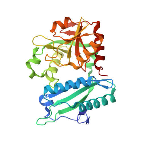 | |
UniProt | |||||
Entity Groups | |||||
| Sequence Clusters | 30% Identity50% Identity70% Identity90% Identity95% Identity100% Identity | ||||
| UniProt Group | Q79FW0 | ||||
Sequence AnnotationsExpand | |||||
Reference Sequence | |||||
| Ligands 3 Unique | |||||
|---|---|---|---|---|---|
| ID | Chains | Name / Formula / InChI Key | 2D Diagram | 3D Interactions | |
| PLP (Subject of Investigation/LOI) Download:Ideal Coordinates CCD File | E [auth A], H [auth B], I [auth C], K [auth D] | PYRIDOXAL-5'-PHOSPHATE C8 H10 N O6 P NGVDGCNFYWLIFO-UHFFFAOYSA-N |  | ||
| CIT Download:Ideal Coordinates CCD File | F [auth A], J [auth C] | CITRIC ACID C6 H8 O7 KRKNYBCHXYNGOX-UHFFFAOYSA-N |  | ||
| GOL Download:Ideal Coordinates CCD File | G [auth A] | GLYCEROL C3 H8 O3 PEDCQBHIVMGVHV-UHFFFAOYSA-N |  | ||
| Length ( Å ) | Angle ( ˚ ) |
|---|---|
| a = 66.431 | α = 90 |
| b = 203.248 | β = 89.96 |
| c = 48.112 | γ = 90 |
| Software Name | Purpose |
|---|---|
| HKL-2000 | data scaling |
| PHENIX | refinement |
| PDB_EXTRACT | data extraction |
| HKL-2000 | data reduction |
| PHASER | phasing |


