6PXL

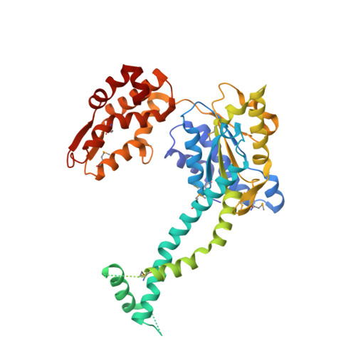
3.74 Angstroms resolution structure of HlsU with an axial-channel plug


Experimental Data Snapshot

  • Method: X-RAY DIFFRACTION
  • Resolution: 3.74 Å
  • R-Value Free: 0.265 
  • R-Value Work: 0.220 
  • R-Value Observed: 0.223 

wwPDB Validation   3D Report Full Report


Ligand Structure Quality Assessment 


This is version 1.3 of the entry. See complete history


Literature

Heat activates the AAA+ HslUV protease by melting an axial autoinhibitory plug.

Baytshtok, V.Fei, X.Shih, T.T.Grant, R.A.Santos, J.C.Baker, T.A.Sauer, R.T.

(2021) Cell Rep 34: 108639-108639

  • DOI: https://doi.org/10.1016/j.celrep.2020.108639
  • Primary Citation of Related Structures:  
    6PXI, 6PXK, 6PXL

  • PubMed Abstract: 

    At low temperatures, protein degradation by the AAA+ HslUV protease is very slow. New crystal structures reveal that residues in the intermediate domain of the HslU 6 unfoldase can plug its axial channel, blocking productive substrate binding and subsequent unfolding, translocation, and degradation by the HslV 12 peptidase. Biochemical experiments with wild-type and mutant enzymes support a model in which heat-induced melting of this autoinhibitory plug activates HslUV proteolysis.


  • Organizational Affiliation

    Department of Biology, Massachusetts Institute of Technology, Cambridge, MA 02139, USA.


Macromolecules
Find similar proteins by:  (by identity cutoff)  |  3D Structure
Entity ID: 1
MoleculeChains Sequence LengthOrganismDetailsImage
ATP-dependent protease ATPase subunit HslU
A, B, C, D, E
A, B, C, D, E, F, G, H, I, J, K, L
448Escherichia coliMutation(s): 0 
Gene Names: hslU
UniProt
Find proteins for P0A6H5 (Escherichia coli (strain K12))
Explore P0A6H5 
Go to UniProtKB:  P0A6H5
Entity Groups  
Sequence Clusters30% Identity50% Identity70% Identity90% Identity95% Identity100% Identity
UniProt GroupP0A6H5
Sequence Annotations
Expand
  • Reference Sequence
Small Molecules
Ligands 3 Unique
IDChains Name / Formula / InChI Key2D Diagram3D Interactions
ADP
Query on ADP

Download Ideal Coordinates CCD File 
AA [auth L]
M [auth A]
P [auth B]
Q [auth C]
R [auth D]
AA [auth L],
M [auth A],
P [auth B],
Q [auth C],
R [auth D],
S [auth E],
T [auth F],
U [auth G],
V [auth H],
W [auth I],
Y [auth J],
Z [auth K]
ADENOSINE-5'-DIPHOSPHATE
C10 H15 N5 O10 P2
XTWYTFMLZFPYCI-KQYNXXCUSA-N
SO4
Query on SO4

Download Ideal Coordinates CCD File 
BA [auth L],
N [auth A]
SULFATE ION
O4 S
QAOWNCQODCNURD-UHFFFAOYSA-L
MG
Query on MG

Download Ideal Coordinates CCD File 
CA [auth L],
O [auth A],
X [auth I]
MAGNESIUM ION
Mg
JLVVSXFLKOJNIY-UHFFFAOYSA-N
Modified Residues  1 Unique
IDChains TypeFormula2D DiagramParent
MSE
Query on MSE
A, B, C, D, E
A, B, C, D, E, F, G, H, I, J, K, L
L-PEPTIDE LINKINGC5 H11 N O2 SeMET
Experimental Data & Validation

Experimental Data

  • Method: X-RAY DIFFRACTION
  • Resolution: 3.74 Å
  • R-Value Free: 0.265 
  • R-Value Work: 0.220 
  • R-Value Observed: 0.223 
  • Space Group: C 1 2 1
Unit Cell:
Length ( Å )Angle ( ˚ )
a = 414.594α = 90
b = 92.331β = 114.37
c = 200.849γ = 90
Software Package:
Software NamePurpose
PHENIXrefinement
PDB_EXTRACTdata extraction
HKL-2000data reduction
HKL-2000data scaling
PHASERphasing

Structure Validation

View Full Validation Report


Ligand Structure Quality Assessment 


Entry History & Funding Information

Deposition Data


Funding OrganizationLocationGrant Number
National Institutes of Health/National Human Genome Research Institute (NIH/NHGRI)United States--

Revision History  (Full details and data files)

  • Version 1.0: 2020-07-29
    Type: Initial release
  • Version 1.1: 2021-02-10
    Changes: Database references
  • Version 1.2: 2023-10-11
    Changes: Data collection, Database references, Refinement description
  • Version 1.3: 2023-11-15
    Changes: Data collection