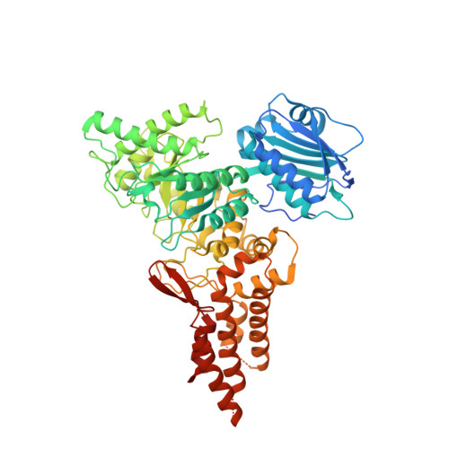
6PV4

Structure of CpGH84A


Experimental Data Snapshot

  • Method: X-RAY DIFFRACTION
  • Resolution: 2.20 Å
  • R-Value Free: 0.248 
  • R-Value Work: 0.202 
  • R-Value Observed: 0.204 

Starting Model: experimental
View more details

wwPDB Validation   3D Report Full Report


This is version 1.4 of the entry. See complete history


Literature

Structural and functional analysis of four family 84 glycoside hydrolases from the opportunistic pathogen Clostridium perfringens.

Pluvinage, B.Massel, P.M.Burak, K.Boraston, A.B.

(2019) Glycobiology 30: 49-57

  • DOI: https://doi.org/10.1093/glycob/cwz069
  • Primary Citation of Related Structures:  
    6PV4, 6PV5, 6PWI

  • PubMed Abstract: 

    The opportunistic pathogen Clostridium perfringens possesses the ability to colonize the protective mucin layer in the gastrointestinal tract. To assist this, the C. perfringens genome contains a battery of genes encoding glycoside hydrolases (GHs) that are likely active on mucin glycans, including four genes encoding family 84 GHs: CpGH84A (NagH), CpGH84B (NagI), CpGH84C (NagJ) and CpGH84D (NagK). To probe the potential advantage gained by the expansion of GH84 enzymes in C. perfringens, we undertook the structural and functional characterization of the CpGH84 catalytic modules. Here, we show that these four CpGH84 catalytic modules act as β-N-acetyl-D-glucosaminidases able to hydrolyze N- and O-glycan motifs. CpGH84A and CpGH84D displayed a substrate specificity restricted to terminal β-1,2- and β-1,6-linked N-acetyl-D-glucosamine (GlcNAc). CpGH84B and CpGH84C appear more promiscuous with activity on terminal β-1,2-, β-1,3- and β-1,6-linked GlcNAc; both possess some activity toward β-1,4-linked GlcNAc, but this is dependent upon which monosaccharide it is linked to. Furthermore, all the CpGH84s have different optimum pHs ranging from 5.2 to 7.0. Consistent with their β-N-acetyl-D-glucosaminidase activities, the structures of the four catalytic modules revealed similar folds with a catalytic site including a conserved -1 subsite that binds GlcNAc. However, nonconserved residues in the vicinity of the +1 subsite suggest different accommodation of the sugar preceding the terminal GlcNAc, resulting in subtly different substrate specificities. This structure-function comparison of the four GH84 catalytic modules from C. perfringens reveals their different biochemical properties, which may relate to how they are deployed in the bacterium's niche in the host.


  • Organizational Affiliation

    Biochemistry and Microbiology, University of Victoria, PO Box 3055 STN CSC, Victoria, BC V8W 3P6, Canada.


Macromolecules
Find similar proteins by:  (by identity cutoff)  |  3D Structure
Entity ID: 1
MoleculeChains Sequence LengthOrganismDetailsImage
Glycoside Hydrolase
A, B, C, D
653Clostridium perfringens ATCC 13124Mutation(s): 0 
Gene Names: nagHCPF_0184
EC: 3.2.1.35
UniProt
Find proteins for A0A0H2YRL1 (Clostridium perfringens (strain ATCC 13124 / DSM 756 / JCM 1290 / NCIMB 6125 / NCTC 8237 / Type A))
Explore A0A0H2YRL1 
Go to UniProtKB:  A0A0H2YRL1
Entity Groups  
Sequence Clusters30% Identity50% Identity70% Identity90% Identity95% Identity100% Identity
UniProt GroupA0A0H2YRL1
Sequence Annotations
Expand
  • Reference Sequence
Small Molecules
Ligands 2 Unique
IDChains Name / Formula / InChI Key2D Diagram3D Interactions
EDO
Query on EDO

Download Ideal Coordinates CCD File 
EA [auth C]
FA [auth C]
GA [auth C]
H [auth A]
HA [auth C]
EA [auth C],
FA [auth C],
GA [auth C],
H [auth A],
HA [auth C],
I [auth A],
IA [auth C],
J [auth A],
JA [auth C],
K [auth A],
KA [auth C],
L [auth A],
M [auth A],
N [auth A],
QA [auth D],
RA [auth D],
SA [auth D],
TA [auth D],
U [auth B],
V [auth B],
W [auth B],
X [auth B],
Y [auth B]
1,2-ETHANEDIOL
C2 H6 O2
LYCAIKOWRPUZTN-UHFFFAOYSA-N
CA
Query on CA

Download Ideal Coordinates CCD File 
AA [auth C]
BA [auth C]
CA [auth C]
DA [auth C]
E [auth A]
AA [auth C],
BA [auth C],
CA [auth C],
DA [auth C],
E [auth A],
F [auth A],
G [auth A],
LA [auth D],
MA [auth D],
NA [auth D],
O [auth B],
OA [auth D],
P [auth B],
PA [auth D],
Q [auth B],
R [auth B],
S [auth B],
T [auth B],
Z [auth C]
CALCIUM ION
Ca
BHPQYMZQTOCNFJ-UHFFFAOYSA-N
Experimental Data & Validation

Experimental Data

  • Method: X-RAY DIFFRACTION
  • Resolution: 2.20 Å
  • R-Value Free: 0.248 
  • R-Value Work: 0.202 
  • R-Value Observed: 0.204 
  • Space Group: P 1
Unit Cell:
Length ( Å )Angle ( ˚ )
a = 92.09α = 95.74
b = 92.11β = 110.52
c = 103.08γ = 119.71
Software Package:
Software NamePurpose
REFMACrefinement
MOSFLMdata reduction
SCALAdata scaling
PHASERphasing
PDB_EXTRACTdata extraction

Structure Validation

View Full Validation Report


Entry History & Funding Information

Deposition Data


Funding OrganizationLocationGrant Number
Canadian Institutes of Health Research (CIHR)Canada--

Revision History  (Full details and data files)

  • Version 1.0: 2019-09-11
    Type: Initial release
  • Version 1.1: 2019-11-20
    Changes: Database references
  • Version 1.2: 2020-01-01
    Changes: Database references
  • Version 1.3: 2020-01-08
    Changes: Author supporting evidence
  • Version 1.4: 2023-10-11
    Changes: Data collection, Database references, Derived calculations, Refinement description