6PSA | pdb_00006psa

PIE12 D-PEPTIDE AGAINST HIV ENTRY (IN COMPLEX WITH IQN17 Q577R RESISTANCE MUTANT)


Experimental Data Snapshot

  • Method: X-RAY DIFFRACTION
  • Resolution: 1.30 Å
  • R-Value Free: 
    0.217 (Depositor), 0.216 (DCC) 
  • R-Value Work: 
    0.176 (Depositor), 0.175 (DCC) 
  • R-Value Observed: 
    0.180 (Depositor) 

wwPDB Validation 3D Report Full Report

Validation slider image for 6PSA

This is version 1.1 of the entry. See complete history

Literature

Characterization of resistance to a potent D-peptide HIV entry inhibitor.

Smith, A.R.Weinstock, M.T.Siglin, A.E.Whitby, F.G.Francis, J.N.Hill, C.P.Eckert, D.M.Root, M.J.Kay, M.S.

(2019) Retrovirology 16: 28-28

  • DOI: https://doi.org/10.1186/s12977-019-0489-7
  • Primary Citation Related Structures: 
    6PSA

  • PubMed Abstract: 

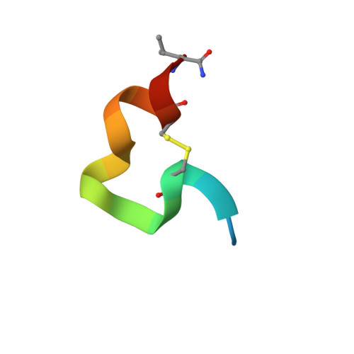
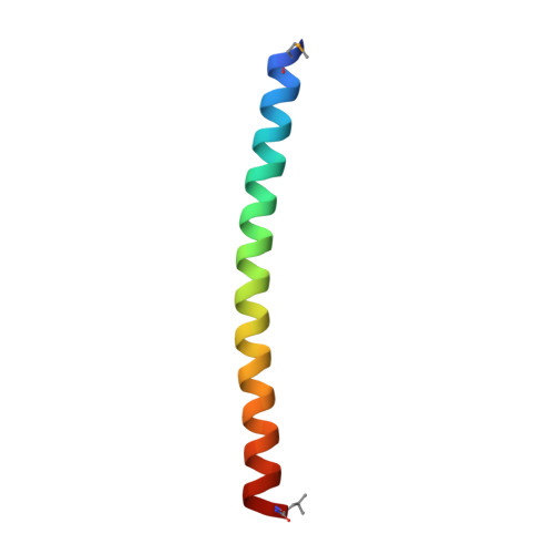
    PIE12-trimer is a highly potent D-peptide HIV-1 entry inhibitor that broadly targets group M isolates. It specifically binds the three identical conserved hydrophobic pockets at the base of the gp41 N-trimer with sub-femtomolar affinity. This extremely high affinity for the transiently exposed gp41 trimer provides a reserve of binding energy (resistance capacitor) to prevent the viral resistance pathway of stepwise accumulation of modest affinity-disrupting mutations. Such modest mutations would not affect PIE12-trimer potency and therefore not confer a selective advantage. Viral passaging in the presence of escalating PIE12-trimer concentrations ultimately selected for PIE12-trimer resistant populations, but required an extremely extended timeframe (> 1 year) in comparison to other entry inhibitors. Eventually, HIV developed resistance to PIE12-trimer by mutating Q577 in the gp41 pocket. Using deep sequence analysis, we identified three mutations at Q577 (R, N and K) in our two PIE12-trimer resistant pools. Each point mutant is capable of conferring the majority of PIE12-trimer resistance seen in the polyclonal pools. Surface plasmon resonance studies demonstrated substantial affinity loss between PIE12-trimer and the Q577R-mutated gp41 pocket. A high-resolution X-ray crystal structure of PIE12 bound to the Q577R pocket revealed the loss of two hydrogen bonds, the repositioning of neighboring residues, and a small decrease in buried surface area. The Q577 mutations in an NL4-3 backbone decreased viral growth rates. Fitness was ultimately rescued in resistant viral pools by a suite of compensatory mutations in gp120 and gp41, of which we identified seven candidates from our sequencing data. These data show that PIE12-trimer exhibits a high barrier to resistance, as extended passaging was required to develop resistant virus with normal growth rates. The primary resistance mutation, Q577R/N/K, found in the conserved gp41 pocket, substantially decreases inhibitor affinity but also damages viral fitness, and candidate compensatory mutations in gp160 have been identified.


  • Organizational Affiliation
    • Department of Biochemistry, University of Utah School of Medicine, Salt Lake City, UT, 84112, USA.

Macromolecule Content 

  • Total Structure Weight: 7.64 kDa 
  • Atom Count: 606 
  • Modeled Residue Count: 65 
  • Deposited Residue Count: 65 
  • Unique protein chains: 2

Macromolecules

Find similar proteins by:  Sequence   |   3D Structure  
Entity ID: 1
MoleculeChains  Sequence LengthOrganismDetailsImage
PIE12 D-peptideA [auth H]18synthetic constructMutation(s): 0 
Find similar proteins by:|  3D Structure
Entity ID: 2
MoleculeChains  Sequence LengthOrganismDetailsImage
IQN17B [auth A]47Saccharomyces cerevisiaeHuman immunodeficiency virus 1
This entity is chimeric
Mutation(s): 3 
UniProt
Find proteins for P03069 (Saccharomyces cerevisiae (strain ATCC 204508 / S288c))
Explore P03069 
Go to UniProtKB:  P03069
Find proteins for P04578 (Human immunodeficiency virus type 1 group M subtype B (isolate HXB2))
Explore P04578 
Go to UniProtKB:  P04578
Entity Groups
Sequence Clusters30% Identity50% Identity70% Identity90% Identity95% Identity100% Identity
UniProt GroupsP03069P04578
Sequence Annotations
Expand
Reference Sequence

Small Molecules

Modified Residues  1 Unique
IDChains TypeFormula2D DiagramParent
DAS
Query on DAS
A [auth H]D-PEPTIDE LINKINGC4 H7 N O4

--

DCY
Query on DCY
A [auth H]D-PEPTIDE LINKINGC3 H7 N O2 S

--

DGL
Query on DGL
A [auth H]D-PEPTIDE LINKINGC5 H9 N O4

--

DGN
Query on DGN
A [auth H]D-PEPTIDE LINKINGC5 H10 N2 O3

--

DHI
Query on DHI
A [auth H]D-PEPTIDE LINKINGC6 H10 N3 O2

--

DLE
Query on DLE
A [auth H]D-PEPTIDE LINKINGC6 H13 N O2

--

DLY
Query on DLY
A [auth H]D-PEPTIDE LINKINGC6 H14 N2 O2

--

DPR
Query on DPR
A [auth H]D-PEPTIDE LINKINGC5 H9 N O2

--

DTR
Query on DTR
A [auth H]D-PEPTIDE LINKINGC11 H12 N2 O2

--

DTY
Query on DTY
A [auth H]D-PEPTIDE LINKINGC9 H11 N O3

--

MSE
Query on MSE
B [auth A]L-PEPTIDE LINKINGC5 H11 N O2 SeMET

Experimental Data & Validation

Experimental Data

  • Method: X-RAY DIFFRACTION
  • Resolution: 1.30 Å
  • R-Value Free:  0.217 (Depositor), 0.216 (DCC) 
  • R-Value Work:  0.176 (Depositor), 0.175 (DCC) 
  • R-Value Observed: 0.180 (Depositor) 
Space Group: H 3
Unit Cell:
Length ( Å )Angle ( ˚ )
a = 48.843α = 90
b = 48.843β = 90
c = 67.634γ = 120
Software Package:
Software NamePurpose
PHENIXrefinement
HKL-2000data reduction
SCALEPACKdata scaling
PHASERphasing

Structure Validation

View Full Validation Report


Entry History 

& Funding Information

Deposition Data


Funding OrganizationLocationGrant Number
National Institutes of Health/National Human Genome Research Institute (NIH/NHGRI)United States--

Revision History  (Full details and data files)

  • Version 1.0: 2020-02-05
    Type: Initial release
  • Version 1.1: 2024-11-20
    Changes: Data collection, Database references, Structure summary