6PNL | pdb_00006pnl

Structure of Epimerase Mth375 from the thermophilic pseudomurein-containing methanogen Methanothermobacter thermautotrophicus


Experimental Data Snapshot

  • Method: X-RAY DIFFRACTION
  • Resolution: 2.01 Å
  • R-Value Free: 
    0.190 (Depositor), 0.194 (DCC) 
  • R-Value Work: 
    0.162 (Depositor), 0.166 (DCC) 
  • R-Value Observed: 
    0.163 (Depositor) 

Starting Model: experimental
View more details

wwPDB Validation 3D Report Full Report

Validation slider image for 6PNL

Ligand Structure Quality Assessment 


This is version 1.5 of the entry. See complete history

Literature

Structural characterisation of nucleotide sugar short-chain dehydrogenases/reductases from the thermophilic pseudomurein-containing methanogen Methanothermobacter thermautotrophicus Delta H.

Carbone, V.Schofield, L.R.Edwards, P.J.B.Sutherland-Smith, A.J.Ronimus, R.S.

(2025) FEBS J 

  • DOI: https://doi.org/10.1111/febs.70248
  • Primary Citation Related Structures: 
    6PMH, 6PNL, 8W3U, 9AR1

  • PubMed Abstract: 

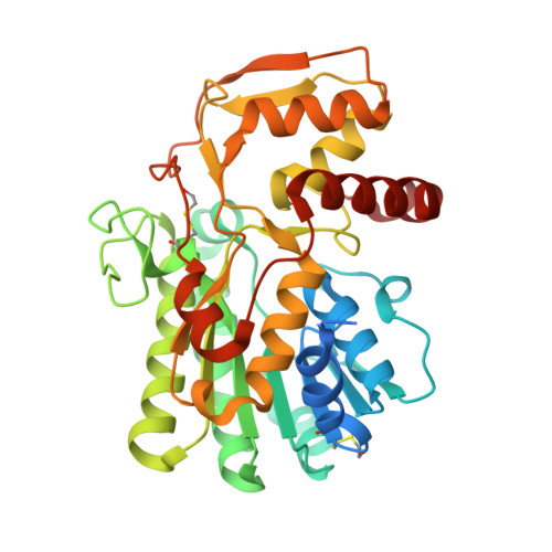
    Epimerases and dehydratases are widely studied members of the extended short-chain dehydrogenase/reductase (SDR) enzyme superfamily and are important in nucleotide sugar conversion and diversification, for example, the interconversion of uridine diphosphate (UDP)-linked glucose and galactose. Methanothermobacter thermautotrophicus contains a cluster of genes, the annotations of which indicate involvement in glycan biosynthesis such as that of cell walls or capsular polysaccharides. In particular, genes encoding UDP-glucose 4-epimerase related protein (Mth375), UDP-glucose 4-epimerase homologue (Mth380) and dTDP-glucose 4,6-dehydratase related protein (Mth373) may be involved in the biosynthesis of an unusual aminosugar in pseudomurein. In this paper, we present the structures of Mth375, an archaeal sugar epimerase/dehydratase protein (WbmF) determined to a resolution of 2.0 Å. The structure contains an N-terminal Rossmann-fold domain with bound nicotinamide adenine dinucleotide hydride (NADH) and a C-terminal catalytic domain with bound UDP. We also present the structure for Mth373 co-crystallised with uridine-5'-diphosphate-xylopyranose to a resolution of 1.96 Å as a NAD + -dependent oxidative decarboxylase (UDP-xylose synthase; EC4.1.1.35). Molecular modelling has also allowed for the identification of Mth380 as a UDP-N-acetylglucosamine 4-epimerase (WbpP; EC5.1.3.7), Mth631 as a UDP-glucose 4-epimerase (GalE; EC5.1.3.2) and Mth1789 as a classical dTDP-d-glucose 4,6-dehydratase (EC4.2.1.46). The UDP-sugar specificity of each archaeal nucleotide sugar short-chain dehydrogenase/reductase (NS-SDR) was elucidated via sequence, molecular modelling and structural analyses. Overall, these structures potentially shed light on the formation of the glycan portion of pseudomurein and capsular polysaccharide in Archaea.


  • Organizational Affiliation
    • AgResearch Ltd., Grasslands, Palmerston North, New Zealand.

Macromolecule Content 

  • Total Structure Weight: 43.4 kDa 
  • Atom Count: 3,087 
  • Modeled Residue Count: 336 
  • Deposited Residue Count: 368 
  • Unique protein chains: 1

Macromolecules

Find similar proteins by:|  3D Structure
Entity ID: 1
MoleculeChains  Sequence LengthOrganismDetailsImage
UDP-glucose 4-epimerase related protein368Methanothermobacter thermautotrophicusMutation(s): 0 
UniProt
Find proteins for O26475 (Methanothermobacter thermautotrophicus (strain ATCC 29096 / DSM 1053 / JCM 10044 / NBRC 100330 / Delta H))
Explore O26475 
Go to UniProtKB:  O26475
Entity Groups
Sequence Clusters30% Identity50% Identity70% Identity90% Identity95% Identity100% Identity
UniProt GroupO26475
Sequence Annotations
Expand
Reference Sequence

Small Molecules

Ligands 4 Unique
IDChains Name / Formula / InChI Key2D Diagram3D Interactions
NAD
(Subject of Investigation/LOI)

Query on NAD


Download:Ideal Coordinates CCD File
B [auth A]NICOTINAMIDE-ADENINE-DINUCLEOTIDE
C21 H27 N7 O14 P2
BAWFJGJZGIEFAR-NNYOXOHSSA-N
UDP
(Subject of Investigation/LOI)

Query on UDP


Download:Ideal Coordinates CCD File
C [auth A]URIDINE-5'-DIPHOSPHATE
C9 H14 N2 O12 P2
XCCTYIAWTASOJW-XVFCMESISA-N
SO4

Query on SO4


Download:Ideal Coordinates CCD File
G [auth A]SULFATE ION
O4 S
QAOWNCQODCNURD-UHFFFAOYSA-L
EDO

Query on EDO


Download:Ideal Coordinates CCD File
D [auth A],
E [auth A],
F [auth A]
1,2-ETHANEDIOL
C2 H6 O2
LYCAIKOWRPUZTN-UHFFFAOYSA-N
Modified Residues  1 Unique
IDChains TypeFormula2D DiagramParent
CME
Query on CME
A
L-PEPTIDE LINKINGC5 H11 N O3 S2CYS

Experimental Data & Validation

Experimental Data

  • Method: X-RAY DIFFRACTION
  • Resolution: 2.01 Å
  • R-Value Free:  0.190 (Depositor), 0.194 (DCC) 
  • R-Value Work:  0.162 (Depositor), 0.166 (DCC) 
  • R-Value Observed: 0.163 (Depositor) 
Space Group: P 63 2 2
Unit Cell:
Length ( Å )Angle ( ˚ )
a = 89.311α = 90
b = 89.311β = 90
c = 174.695γ = 120
Software Package:
Software NamePurpose
XDSdata reduction
Aimlessdata scaling
MOLREPphasing
REFMACrefinement
PDB_EXTRACTdata extraction

Structure Validation

View Full Validation Report


Ligand Structure Quality Assessment 


Entry History 

& Funding Information

Deposition Data


Funding OrganizationLocationGrant Number
Royal Society of New ZealandNew ZealandAGR1301

Revision History  (Full details and data files)

  • Version 1.0: 2020-07-08
    Type: Initial release
  • Version 1.1: 2023-08-09
    Changes: Database references, Source and taxonomy
  • Version 1.2: 2023-10-11
    Changes: Data collection, Refinement description
  • Version 1.3: 2023-12-27
    Changes: Structure summary
  • Version 1.4: 2024-10-23
    Changes: Structure summary
  • Version 1.5: 2025-09-17
    Changes: Database references