6PFT | pdb_00006pft

rsEGFP2 with a chlorinated chromophore in the non-fluorescent off-state


Experimental Data Snapshot

  • Method: X-RAY DIFFRACTION
  • Resolution: 1.45 Å
  • R-Value Free: 
    0.176 (Depositor), 0.183 (DCC) 
  • R-Value Work: 
    0.150 (Depositor), 0.162 (DCC) 
  • R-Value Observed: 
    0.151 (Depositor) 

Starting Model: experimental
View more details

wwPDB Validation 3D Report Full Report

Validation slider image for 6PFT

This is version 3.0 of the entry. See complete history

Literature

Structural Evidence of Photoisomerization Pathways in Fluorescent Proteins.

Chang, J.Romei, M.G.Boxer, S.G.

(2019) J Am Chem Soc 141: 15504-15508

  • DOI: https://doi.org/10.1021/jacs.9b08356
  • Primary Citation Related Structures: 
    6PFR, 6PFS, 6PFT, 6PFU

  • PubMed Abstract: 

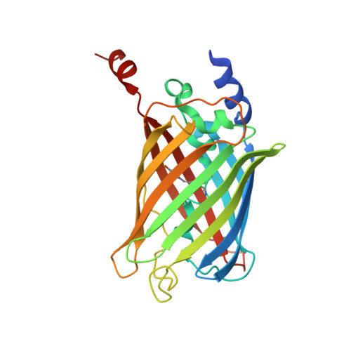
    Double-bond photoisomerization in molecules such as the green fluorescent protein (GFP) chromophore can occur either via a volume-demanding one-bond-flip pathway or via a volume-conserving hula-twist pathway. Understanding the factors that determine the pathway of photoisomerization would inform the rational design of photoswitchable GFPs as improved tools for super-resolution microscopy. In this communication, we reveal the photoisomerization pathway of a photoswitchable GFP, rsEGFP2, by solving crystal structures of cis and trans rsEGFP2 containing a monochlorinated chromophore. The position of the chlorine substituent in the trans state breaks the symmetry of the phenolate ring of the chromophore and allows us to distinguish the two pathways. Surprisingly, we find that the pathway depends on the arrangement of protein monomers within the crystal lattice: in a looser packing, the one-bond-flip occurs, whereas, in a tighter packing (7% smaller unit cell size), the hula-twist occurs.


  • Organizational Affiliation
    • Department of Physics , Stanford University , Stanford , California 94305 , United States.

Macromolecule Content 

  • Total Structure Weight: 28.66 kDa 
  • Atom Count: 2,316 
  • Modeled Residue Count: 241 
  • Deposited Residue Count: 250 
  • Unique protein chains: 1

Macromolecules

Find similar proteins by:|  3D Structure
Entity ID: 1
MoleculeChains  Sequence LengthOrganismDetailsImage
Green fluorescent protein250Aequorea victoriaMutation(s): 0 
UniProt
Find proteins for P42212 (Aequorea victoria)
Explore P42212 
Go to UniProtKB:  P42212
Entity Groups
Sequence Clusters30% Identity50% Identity70% Identity90% Identity95% Identity100% Identity
UniProt GroupP42212
Sequence Annotations
Expand
Reference Sequence

Small Molecules

Ligands 1 Unique
IDChains Name / Formula / InChI Key2D Diagram3D Interactions
SO4

Query on SO4


Download:Ideal Coordinates CCD File
B [auth A]SULFATE ION
O4 S
QAOWNCQODCNURD-UHFFFAOYSA-L
Modified Residues  1 Unique
IDChains TypeFormula2D DiagramParent
OHD
Query on OHD
A
L-PEPTIDE LINKINGC14 H14 Cl N3 O4ALA, TYR, GLY

Experimental Data & Validation

Experimental Data

  • Method: X-RAY DIFFRACTION
  • Resolution: 1.45 Å
  • R-Value Free:  0.176 (Depositor), 0.183 (DCC) 
  • R-Value Work:  0.150 (Depositor), 0.162 (DCC) 
  • R-Value Observed: 0.151 (Depositor) 
Space Group: P 21 21 21
Unit Cell:
Length ( Å )Angle ( ˚ )
a = 51.014α = 90
b = 62.716β = 90
c = 68.619γ = 90
Software Package:
Software NamePurpose
PHENIXrefinement
XDSdata reduction
XDSdata scaling
PHENIXphasing

Structure Validation

View Full Validation Report


Entry History 

& Funding Information

Deposition Data


Funding OrganizationLocationGrant Number
National Institutes of Health/National Institute of General Medical Sciences (NIH/NIGMS)United StatesGM118044

Revision History  (Full details and data files)

  • Version 1.0: 2019-08-07
    Type: Initial release
  • Version 1.1: 2019-10-02
    Changes: Data collection, Database references
  • Version 1.2: 2019-10-16
    Changes: Data collection, Database references
  • Version 2.0: 2020-01-01
    Changes: Author supporting evidence, Polymer sequence, Structure summary
  • Version 2.1: 2023-10-11
    Changes: Data collection, Database references, Refinement description
  • Version 2.2: 2023-11-15
    Changes: Data collection
  • Version 3.0: 2026-03-18
    Changes: Polymer sequence, Structure summary