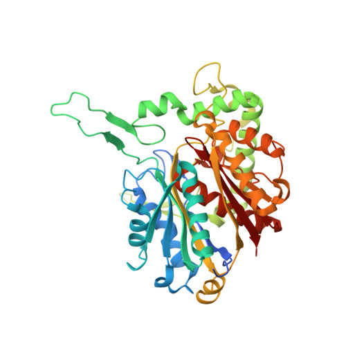
6PCB

Crystal structure of beta-ketoadipyl-CoA thiolase mutant (H356A) in complex with COA


Experimental Data Snapshot

  • Method: X-RAY DIFFRACTION
  • Resolution: 1.61 Å
  • R-Value Free: 0.196 
  • R-Value Work: 0.161 
  • R-Value Observed: 0.161 

Starting Model: experimental
View more details

wwPDB Validation   3D Report Full Report


Ligand Structure Quality Assessment 


This is version 1.3 of the entry. See complete history


Literature

Structural basis for differentiation between two classes of thiolase: Degradative vs biosynthetic thiolase.

Bhaskar, S.Steer, D.L.Anand, R.Panjikar, S.

(2020) J Struct Biol X 4: 100018-100018

  • DOI: https://doi.org/10.1016/j.yjsbx.2019.100018
  • Primary Citation of Related Structures:  
    6PCA, 6PCB, 6PCC, 6PCD

  • PubMed Abstract: 

    Thiolases are a well characterized family of enzymes with two distinct categories: degradative, β-ketoadipyl-CoA thiolases and biosynthetic, acetoacetyl-CoA thiolases. Both classes share an identical catalytic triad but catalyze reactions in opposite directions. Moreover, it is established that in contrast to the biosynthetic thiolases the degradative thiolases can accept substrates with broad chain lengths. Hitherto, no residue or structural pattern has been recognized that might help to discern the two thiolases, here we exploit, a tetrameric degradative thiolase from Pseudomonas putida KT2440 annotated as PcaF, as a model system to understand features which distinguishes the two classes using structural studies and bioinformatics analyses. Degradative thiolases have different active site architecture when compared to biosynthetic thiolases, demonstrating the dissimilar chemical nature of the active site architecture. Both thiolases deploy different "anchoring residues" to tether the large Coenzyme A (CoA) or CoA derivatives. Interestingly, the H356 of the catalytic triad in PcaF is directly involved in tethering the CoA/CoA derivatives into the active site and we were able to trap a gridlocked thiolase structure of the H356A mutant, where the CoA was found to be covalently linked to the catalytic cysteine residue, inhibiting the overall reaction. Further, X-ray structures with two long chain CoA derivatives, hexanal-CoA and octanal-CoA helped in delineating the long tunnel of 235 Å 2 surface area in PcaF and led to identification of a unique covering loop exclusive to degradative thiolases that plays an active role in determining the tunnel length and the nature of the binding substrate.


  • Organizational Affiliation

    IITB-Monash Research Academy, Mumbai 400076, Maharashtra, India.


Macromolecules
Find similar proteins by:  (by identity cutoff)  |  3D Structure
Entity ID: 1
MoleculeChains Sequence LengthOrganismDetailsImage
Beta-ketoadipyl-CoA thiolase
A, B, C, D
423Pseudomonas putida KT2440Mutation(s): 1 
Gene Names: pcaF-IPP_1377
EC: 2.3.1.16 (PDB Primary Data), 2.3.1.174 (PDB Primary Data)
UniProt
Find proteins for Q88N39 (Pseudomonas putida (strain ATCC 47054 / DSM 6125 / CFBP 8728 / NCIMB 11950 / KT2440))
Explore Q88N39 
Go to UniProtKB:  Q88N39
Entity Groups  
Sequence Clusters30% Identity50% Identity70% Identity90% Identity95% Identity100% Identity
UniProt GroupQ88N39
Sequence Annotations
Expand
  • Reference Sequence
Small Molecules
Ligands 3 Unique
IDChains Name / Formula / InChI Key2D Diagram3D Interactions
COA (Subject of Investigation/LOI)
Query on COA

Download Ideal Coordinates CCD File 
E [auth A],
H [auth B],
K [auth C],
N [auth D]
COENZYME A
C21 H36 N7 O16 P3 S
RGJOEKWQDUBAIZ-IBOSZNHHSA-N
GOL (Subject of Investigation/LOI)
Query on GOL

Download Ideal Coordinates CCD File 
F [auth A],
I [auth B],
L [auth C],
O [auth D]
GLYCEROL
C3 H8 O3
PEDCQBHIVMGVHV-UHFFFAOYSA-N
CL (Subject of Investigation/LOI)
Query on CL

Download Ideal Coordinates CCD File 
G [auth A],
J [auth B],
M [auth C],
P [auth D],
Q [auth D]
CHLORIDE ION
Cl
VEXZGXHMUGYJMC-UHFFFAOYSA-M
Modified Residues  1 Unique
IDChains TypeFormula2D DiagramParent
CSO
Query on CSO
A, B, C, D
L-PEPTIDE LINKINGC3 H7 N O3 SCYS
Experimental Data & Validation

Experimental Data

  • Method: X-RAY DIFFRACTION
  • Resolution: 1.61 Å
  • R-Value Free: 0.196 
  • R-Value Work: 0.161 
  • R-Value Observed: 0.161 
  • Space Group: P 21 21 21
Unit Cell:
Length ( Å )Angle ( ˚ )
a = 110.487α = 90
b = 115.958β = 90
c = 128.866γ = 90
Software Package:
Software NamePurpose
REFMACrefinement
XDSdata reduction
Aimlessdata scaling
Auto-Rickshawphasing

Structure Validation

View Full Validation Report


Ligand Structure Quality Assessment 


Entry History 

Deposition Data

Revision History  (Full details and data files)

  • Version 1.0: 2020-05-27
    Type: Initial release
  • Version 1.1: 2020-07-22
    Changes: Database references
  • Version 1.2: 2024-04-03
    Changes: Data collection, Database references, Refinement description
  • Version 1.3: 2024-11-06
    Changes: Structure summary