6P7M | pdb_00006p7m

Cryo-EM structure of LbCas12a-crRNA: AcrVA4 (1:2 complex)


Experimental Data Snapshot

  • Method: ELECTRON MICROSCOPY
  • Resolution: 3.00 Å
  • Aggregation State: PARTICLE 
  • Reconstruction Method: SINGLE PARTICLE 

wwPDB Validation 3D Report Full Report

Validation slider image for 6P7M

This is version 1.3 of the entry. See complete history

Literature

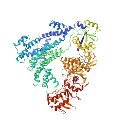
Structural basis for AcrVA4 inhibition of specific CRISPR-Cas12a.

Knott, G.J.Cress, B.F.Liu, J.J.Thornton, B.W.Lew, R.J.Al-Shayeb, B.Rosenberg, D.J.Hammel, M.Adler, B.A.Lobba, M.J.Xu, M.Arkin, A.P.Fellmann, C.Doudna, J.A.

(2019) Elife 8

  • DOI: https://doi.org/10.7554/eLife.49110
  • Primary Citation Related Structures: 
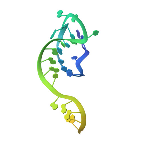
    6P7M, 6P7N

  • PubMed Abstract: 

    CRISPR-Cas systems provide bacteria and archaea with programmable immunity against mobile genetic elements. Evolutionary pressure by CRISPR-Cas has driven bacteriophage to evolve small protein inhibitors, anti-CRISPRs (Acrs), that block Cas enzyme function by wide-ranging mechanisms. We show here that the inhibitor AcrVA4 uses a previously undescribed strategy to recognize the L. bacterium Cas12a (LbCas12a) pre-crRNA processing nuclease, forming a Cas12a dimer, and allosterically inhibiting DNA binding. The Ac. species Cas12a (AsCas12a) enzyme, widely used for genome editing applications, contains an ancestral helical bundle that blocks AcrVA4 binding and allows it to escape anti-CRISPR recognition. Using biochemical, microbiological, and human cell editing experiments, we show that Cas12a orthologs can be rendered either sensitive or resistant to AcrVA4 through rational structural engineering informed by evolution. Together, these findings explain a new mode of CRISPR-Cas inhibition and illustrate how structural variability in Cas effectors can drive opportunistic co-evolution of inhibitors by bacteriophage.


  • Organizational Affiliation
    • Department of Molecular and Cell Biology, University of California, Berkeley, Berkeley, United States.

Macromolecule Content 

  • Total Structure Weight: 184.67 kDa 
  • Atom Count: 10,141 
  • Modeled Residue Count: 1,194 
  • Deposited Residue Count: 1,508 
  • Unique protein chains: 2
  • Unique nucleic acid chains: 1

Macromolecules


Find similar proteins by:|  3D Structure
Entity ID: 1
MoleculeChains  Sequence LengthOrganismDetailsImage
Cas12a1,231Lachnospiraceae bacterium ND2006Mutation(s): 0 
Gene Names: lbcas12a
UniProt
Find proteins for A0ACD6BAZ6 (Lachnospiraceae bacterium ND2006)
Explore A0ACD6BAZ6 
Go to UniProtKB:  A0ACD6BAZ6
Entity Groups
Sequence Clusters30% Identity50% Identity70% Identity90% Identity95% Identity100% Identity
UniProt GroupA0ACD6BAZ6
Sequence Annotations
Expand
Reference Sequence
Find similar proteins by:|  3D Structure
Entity ID: 3
MoleculeChains  Sequence LengthOrganismDetailsImage
anti-CRISPR VA4237Moraxella bovoculiMutation(s): 0 
Gene Names: AAX07_09545
Find similar nucleic acids by:  (by identity cutoff) 
Entity ID: 2
MoleculeChains LengthOrganismImage
mature crRNA40Lachnospiraceae bacterium ND2006
Sequence Annotations
Expand
Reference Sequence

Experimental Data & Validation

Experimental Data

  • Method: ELECTRON MICROSCOPY
  • Resolution: 3.00 Å
  • Aggregation State: PARTICLE 
  • Reconstruction Method: SINGLE PARTICLE 

Structure Validation

View Full Validation Report


Entry History 

& Funding Information

Deposition Data


Funding OrganizationLocationGrant Number
Howard Hughes Medical Institute (HHMI)United States--

Revision History  (Full details and data files)

  • Version 1.0: 2019-08-21
    Type: Initial release
  • Version 1.1: 2019-11-20
    Changes: Author supporting evidence
  • Version 1.2: 2019-12-18
    Changes: Other
  • Version 1.3: 2024-03-20
    Changes: Data collection, Database references