6P4C | pdb_00006p4c

HyHEL10 Fab carrying four heavy chain mutations (HyHEL10-4x): L4F, Y33H, S56N, and Y58F


Experimental Data Snapshot

  • Method: X-RAY DIFFRACTION
  • Resolution: 1.85 Å
  • R-Value Free: 
    0.223 (Depositor), 0.229 (DCC) 
  • R-Value Work: 
    0.188 (Depositor), 0.195 (DCC) 
  • R-Value Observed: 
    0.190 (Depositor) 

Starting Model: experimental
View more details

wwPDB Validation 3D Report Full Report

Validation slider image for 6P4C

This is version 1.3 of the entry. See complete history

Literature

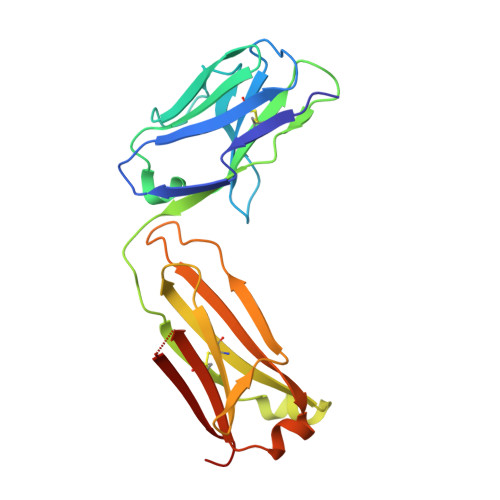
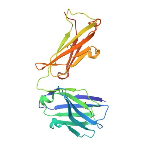
Conformational diversity facilitates antibody mutation trajectories and discrimination between foreign and self-antigens.

Burnett, D.L.Schofield, P.Langley, D.B.Jackson, J.Bourne, K.Wilson, E.Porebski, B.T.Buckle, A.M.Brink, R.Goodnow, C.C.Christ, D.

(2020) Proc Natl Acad Sci U S A 117: 22341-22350

  • DOI: https://doi.org/10.1073/pnas.2005102117
  • Primary Citation Related Structures: 
    6P4A, 6P4B, 6P4C, 6P4D

  • PubMed Abstract: 

    Conformational diversity and self-cross-reactivity of antigens have been correlated with evasion from neutralizing antibody responses. We utilized single cell B cell sequencing, biolayer interferometry and X-ray crystallography to trace mutation selection pathways where the antibody response must resolve cross-reactivity between foreign and self-proteins bearing near-identical contact surfaces, but differing in conformational flexibility. Recurring antibody mutation trajectories mediate long-range rearrangements of framework (FW) and complementarity determining regions (CDRs) that increase binding site conformational diversity. These antibody mutations decrease affinity for self-antigen 19-fold and increase foreign affinity 67-fold, to yield a more than 1,250-fold increase in binding discrimination. These results demonstrate how conformational diversity in antigen and antibody does not act as a barrier, as previously suggested, but rather facilitates high affinity and high discrimination between foreign and self.


  • Organizational Affiliation
    • Garvan Institute of Medical Research, University of New South Wales (UNSW) Sydney, Darlinghurst, NSW 2010, Australia.

Macromolecule Content 

  • Total Structure Weight: 48.15 kDa 
  • Atom Count: 3,367 
  • Modeled Residue Count: 420 
  • Deposited Residue Count: 437 
  • Unique protein chains: 2

Macromolecules

Find similar proteins by:|  3D Structure
Entity ID: 1
MoleculeChains  Sequence LengthOrganismDetailsImage
HyHEL10 Fab light chainA [auth L]214Mus musculusMutation(s): 0 
UniProt
Find proteins for A0A0E4B213 (Mus musculus)
Explore A0A0E4B213 
Go to UniProtKB:  A0A0E4B213
Entity Groups
Sequence Clusters30% Identity50% Identity70% Identity90% Identity95% Identity100% Identity
UniProt GroupA0A0E4B213
Sequence Annotations
Expand
Reference Sequence
Find similar proteins by:|  3D Structure
Entity ID: 2
MoleculeChains  Sequence LengthOrganismDetailsImage
HyHEL10 Fab heavy chainB [auth H]223Mus musculusMutation(s): 0 

Experimental Data & Validation

Experimental Data

  • Method: X-RAY DIFFRACTION
  • Resolution: 1.85 Å
  • R-Value Free:  0.223 (Depositor), 0.229 (DCC) 
  • R-Value Work:  0.188 (Depositor), 0.195 (DCC) 
  • R-Value Observed: 0.190 (Depositor) 
Space Group: P 1 2 1
Unit Cell:
Length ( Å )Angle ( ˚ )
a = 68.556α = 90
b = 40.156β = 107.57
c = 93.649γ = 90
Software Package:
Software NamePurpose
XDSdata reduction
Aimlessdata scaling
PHASERphasing
REFMACrefinement
PDB_EXTRACTdata extraction

Structure Validation

View Full Validation Report


Entry History 

Deposition Data

Revision History  (Full details and data files)

  • Version 1.0: 2020-05-27
    Type: Initial release
  • Version 1.1: 2020-12-16
    Changes: Database references
  • Version 1.2: 2023-10-11
    Changes: Data collection, Database references, Refinement description
  • Version 1.3: 2024-11-06
    Changes: Structure summary