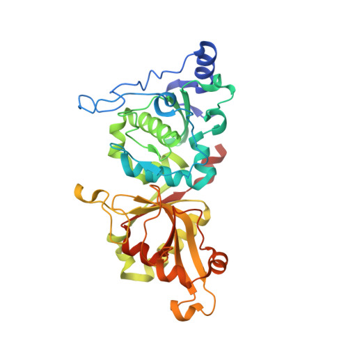
6P3Q

Calpain-5 (CAPN5) Protease Core (PC)


Experimental Data Snapshot

  • Method: X-RAY DIFFRACTION
  • Resolution: 2.80 Å
  • R-Value Free: 0.285 
  • R-Value Work: 0.227 
  • R-Value Observed: 0.230 

Starting Model: in silico
View more details

wwPDB Validation   3D Report Full Report


This is version 1.2 of the entry. See complete history


Literature

Structural Insights into the Unique Activation Mechanisms of a Non-classical Calpain and Its Disease-Causing Variants.

Velez, G.Sun, Y.J.Khan, S.Yang, J.Herrmann, J.Chemudupati, T.MacLaren, R.E.Gakhar, L.Wakatsuki, S.Bassuk, A.G.Mahajan, V.B.

(2020) Cell Rep 30: 881-892.e5

  • DOI: https://doi.org/10.1016/j.celrep.2019.12.077
  • Primary Citation of Related Structures:  
    6P3Q

  • PubMed Abstract: 

    Increased calpain activity is linked to neuroinflammation including a heritable retinal disease caused by hyper-activating mutations in the calcium-activated calpain-5 (CAPN5) protease. Although structures for classical calpains are known, the structure of CAPN5, a non-classical calpain, remains undetermined. Here we report the 2.8 Å crystal structure of the human CAPN5 protease core (CAPN5-PC). Compared to classical calpains, CAPN5-PC requires high calcium concentrations for maximal activity. Structure-based phylogenetic analysis and multiple sequence alignment reveal that CAPN5-PC contains three elongated flexible loops compared to its classical counterparts. The presence of a disease-causing mutation (c.799G>A, p.Gly267Ser) on the unique PC2L2 loop reveals a function in this region for regulating enzymatic activity. This mechanism could be transferred to distant calpains, using synthetic calpain hybrids, suggesting an evolutionary mechanism for fine-tuning calpain function by modifying flexible loops. Further, the open (inactive) conformation of CAPN5-PC provides structural insight into CAPN5-specific residues that can guide inhibitor design.


  • Organizational Affiliation

    Omics Laboratory, Department of Ophthalmology, Byers Eye Institute, Stanford University, Palo Alto, CA 94304, USA; Medical Scientist Training Program, University of Iowa, Iowa City, IA 52242, USA.


Macromolecules
Find similar proteins by:  (by identity cutoff)  |  3D Structure
Entity ID: 1
MoleculeChains Sequence LengthOrganismDetailsImage
Calpain-5
A, B
359Homo sapiensMutation(s): 0 
Gene Names: CAPN5NCL3
EC: 3.4.22
UniProt & NIH Common Fund Data Resources
Find proteins for O15484 (Homo sapiens)
Explore O15484 
Go to UniProtKB:  O15484
PHAROS:  O15484
GTEx:  ENSG00000149260 
Entity Groups  
Sequence Clusters30% Identity50% Identity70% Identity90% Identity95% Identity100% Identity
UniProt GroupO15484
Sequence Annotations
Expand
  • Reference Sequence
Experimental Data & Validation

Experimental Data

  • Method: X-RAY DIFFRACTION
  • Resolution: 2.80 Å
  • R-Value Free: 0.285 
  • R-Value Work: 0.227 
  • R-Value Observed: 0.230 
  • Space Group: P 1 21 1
Unit Cell:
Length ( Å )Angle ( ˚ )
a = 84.038α = 90
b = 51.576β = 110.425
c = 110.635γ = 90
Software Package:
Software NamePurpose
PHENIXrefinement
Blu-Icedata collection
XDSdata reduction
Aimlessdata scaling
Cootmodel building
PHASERphasing

Structure Validation

View Full Validation Report


Entry History & Funding Information

Deposition Data


Funding OrganizationLocationGrant Number
National Institutes of Health/National Eye Institute (NIH/NEI)United StatesR01EY024665

Revision History  (Full details and data files)

  • Version 1.0: 2020-02-05
    Type: Initial release
  • Version 1.1: 2024-03-13
    Changes: Data collection, Database references, Refinement description
  • Version 1.2: 2024-04-03
    Changes: Refinement description