


Crystal Structure Analysis of TAF1 Bromodomain
Seo, H.-S., Dhe-Paganon, S.To be published.
Experimental Data Snapshot
Starting Model: experimental
View more details
Entity ID: 1 | |||||
|---|---|---|---|---|---|
| Molecule | Chains | Sequence Length | Organism | Details | Image |
| Transcription initiation factor TFIID subunit 1 | 137 | Homo sapiens | Mutation(s): 0 Gene Names: TAF1, BA2R, CCG1, CCGS, TAF2A EC: 2.3.1.48 (PDB Primary Data), 2.7.11.1 (PDB Primary Data) | 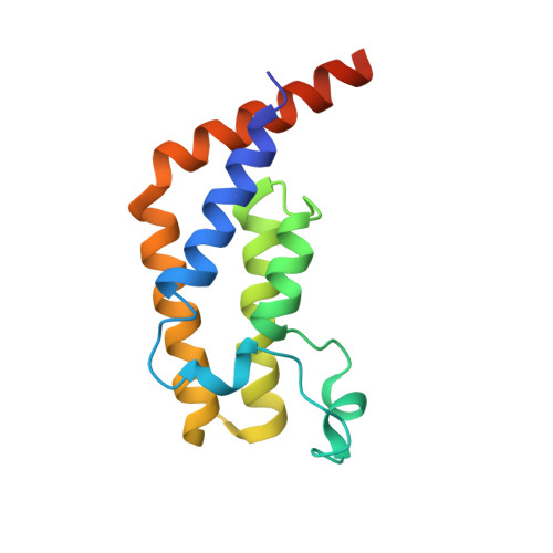 | |
UniProt & NIH Common Fund Data Resources | |||||
PHAROS: P21675 GTEx: ENSG00000147133 | |||||
Entity Groups | |||||
| Sequence Clusters | 30% Identity50% Identity70% Identity90% Identity95% Identity100% Identity | ||||
| UniProt Group | P21675 | ||||
Sequence AnnotationsExpand | |||||
Reference Sequence | |||||
| Ligands 1 Unique | |||||
|---|---|---|---|---|---|
| ID | Chains | Name / Formula / InChI Key | 2D Diagram | 3D Interactions | |
| NQP (Subject of Investigation/LOI) Download:Ideal Coordinates CCD File | C [auth A], D [auth B] | 4-{[(3R)-1-(but-3-en-1-yl)-3-methyl-4-(oxan-4-yl)-2-oxo-1,2,3,4-tetrahydropyrido[2,3-b]pyrazin-6-yl]amino}-3-methoxy-N-(1-methylpiperidin-4-yl)benzamide C31 H42 N6 O4 YEZKKIMJJAQTOO-OAQYLSRUSA-N |  | ||
| Length ( Å ) | Angle ( ˚ ) |
|---|---|
| a = 92.43 | α = 90 |
| b = 92.43 | β = 90 |
| c = 117.43 | γ = 90 |
| Software Name | Purpose |
|---|---|
| XDS | data reduction |
| xia2 | data scaling |
| PHENIX | refinement |
| PDB_EXTRACT | data extraction |
| PHASER | phasing |
| Funding Organization | Location | Grant Number |
|---|---|---|
| National Institutes of Health/National Cancer Institute (NIH/NCI) | United States | -- |


