


Crystal Structure of KDPG Aldolase from Legionella Pneumophila with pyruvate captured at low pH as a covalent carbinolamine intermediate
Davies, D.R., Dranow, D.M.To be published.
Experimental Data Snapshot
Starting Model: experimental
View more details
Entity ID: 1 | |||||
|---|---|---|---|---|---|
| Molecule | Chains | Sequence Length | Organism | Details | Image |
| Keto-deoxy-phosphogluconate aldolase | 228 | Legionella pneumophila | Mutation(s): 0 Gene Names: C3927_01390 EC: 4.1.2.14 (PDB Primary Data), 4.1.3.16 (PDB Primary Data) | 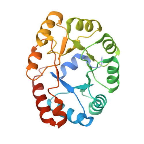 | |
UniProt | |||||
Entity Groups | |||||
| Sequence Clusters | 30% Identity50% Identity70% Identity90% Identity95% Identity100% Identity | ||||
| UniProt Group | Q5ZYF2 | ||||
Sequence AnnotationsExpand | |||||
Reference Sequence | |||||
| Ligands 3 Unique | |||||
|---|---|---|---|---|---|
| ID | Chains | Name / Formula / InChI Key | 2D Diagram | 3D Interactions | |
| PVO (Subject of Investigation/LOI) Download:Ideal Coordinates CCD File | D [auth A], I [auth B], M [auth C] | 2,2-bis(oxidanyl)propanoic acid C3 H6 O4 HPQUMJNDQVOTAZ-UHFFFAOYSA-N |  | ||
| SO4 Download:Ideal Coordinates CCD File | F [auth A] G [auth A] H [auth A] L [auth B] O [auth C] | SULFATE ION O4 S QAOWNCQODCNURD-UHFFFAOYSA-L |  | ||
| EDO Download:Ideal Coordinates CCD File | E [auth A], J [auth B], K [auth B], N [auth C] | 1,2-ETHANEDIOL C2 H6 O2 LYCAIKOWRPUZTN-UHFFFAOYSA-N |  | ||
| Length ( Å ) | Angle ( ˚ ) |
|---|---|
| a = 69.42 | α = 90 |
| b = 79.14 | β = 117.4 |
| c = 70.06 | γ = 90 |
| Software Name | Purpose |
|---|---|
| PHENIX | refinement |
| XSCALE | data scaling |
| XDS | data reduction |
| PHASER | phasing |
| Funding Organization | Location | Grant Number |
|---|---|---|
| National Institutes of Health/National Institute Of Allergy and Infectious Diseases (NIH/NIAID) | United States | -- |


