6OOF | pdb_00006oof

CTX-M-14 Beta Lactamase with Compound 20


Experimental Data Snapshot

  • Method: X-RAY DIFFRACTION
  • Resolution: 1.24 Å
  • R-Value Free: 
    0.201 (Depositor), 0.201 (DCC) 
  • R-Value Work: 
    0.171 (Depositor), 0.172 (DCC) 
  • R-Value Observed: 
    0.171 (Depositor) 

Starting Model: experimental
View more details

wwPDB Validation 3D Report Full Report

Validation slider image for 6OOF

Ligand Structure Quality Assessment 


This is version 1.3 of the entry. See complete history

Literature

An Empirical Study of Amide-Heteroarene pi-Stacking Interactions Using Reversible Inhibitors of a Bacterial Serine Hydrolase.

DeFrees, K.Kemp, M.T.ElHilali-Pollard, X.Zhang, X.Mohamed, A.Chen, Y.Renslo, A.R.

(2019) Org Chem Front 6: 1749-1756

  • DOI: https://doi.org/10.1039/c9qo00342h
  • Primary Citation Related Structures: 
    6OOE, 6OOF, 6OOH, 6OOJ, 6OOK

  • PubMed Abstract: 

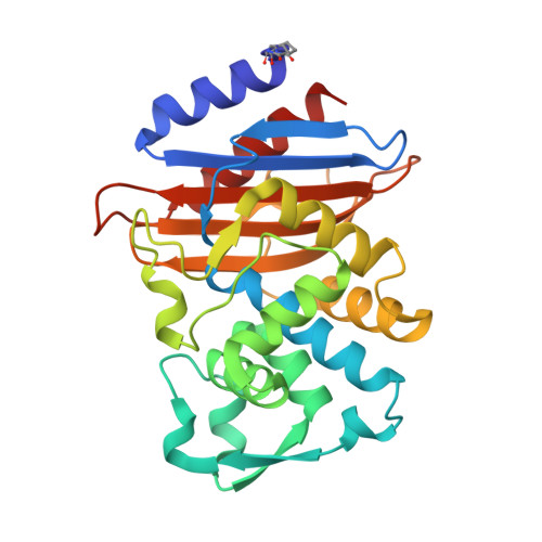
    Compared to aryl-aryl π-stacking interactions, the analogous stacking of heteroarenes on amide π systems is less well understood and vastly underutilized in structure-based drug design. Recent theoretical studies have delineated the important geometric coordinates of the interaction, some of which have been confirmed with synthetic model systems based on Rebek imides. Unfortunately, a broadly useful and tractable protein-ligand model system of this interaction has remained elusive. Here we employed a known inhibitor scaffold to study π-stacking of diverse heteroarene substituents on the amide face of Gly238 in the cephalosporinases CTX-M-14 and CTX-M-27. Biochemical inhibition constants ( K i ) and biophysical binding constants ( K d ) were determined for nineteen new analogues against both enzymes, while multiple high-resolution co-crystal structures revealed remarkably consistent placement of the probe heteroarene on Gly238. The data presented support the predicted importance of opposing dipoles in amide-heteroarene interactions and should be useful for evaluating other theoretical predictions concerning these interactions.


  • Organizational Affiliation
    • Department of Pharmaceutical Chemistry, University of California San Francisco, 600 16th St., San Francisco, California 94158, United States.

Macromolecule Content 

  • Total Structure Weight: 57.57 kDa 
  • Atom Count: 4,982 
  • Modeled Residue Count: 523 
  • Deposited Residue Count: 526 
  • Unique protein chains: 1

Macromolecules

Find similar proteins by:|  3D Structure
Entity ID: 1
MoleculeChains  Sequence LengthOrganismDetailsImage
Beta-lactamase
A, B
263Escherichia coliMutation(s): 0 
Gene Names: blaCTX-M-14
EC: 3.5.2.6
UniProt
Find proteins for Q9L5C7 (Escherichia coli)
Explore Q9L5C7 
Go to UniProtKB:  Q9L5C7
Entity Groups
Sequence Clusters30% Identity50% Identity70% Identity90% Identity95% Identity100% Identity
UniProt GroupQ9L5C7
Sequence Annotations
Expand
Reference Sequence

Experimental Data & Validation

Experimental Data

  • Method: X-RAY DIFFRACTION
  • Resolution: 1.24 Å
  • R-Value Free:  0.201 (Depositor), 0.201 (DCC) 
  • R-Value Work:  0.171 (Depositor), 0.172 (DCC) 
  • R-Value Observed: 0.171 (Depositor) 
Space Group: P 1 21 1
Unit Cell:
Length ( Å )Angle ( ˚ )
a = 45.161α = 90
b = 107.211β = 101.66
c = 47.884γ = 90
Software Package:
Software NamePurpose
PHENIXrefinement
SCALEPACKdata scaling
PDB_EXTRACTdata extraction
HKL-2000data reduction
PHASERphasing

Structure Validation

View Full Validation Report


Ligand Structure Quality Assessment 


Entry History 

& Funding Information

Deposition Data


Funding OrganizationLocationGrant Number
National Institutes of Health/National Institute Of Allergy and Infectious Diseases (NIH/NIAID)United StatesAI103158

Revision History  (Full details and data files)

  • Version 1.0: 2020-07-01
    Type: Initial release
  • Version 1.1: 2020-08-26
    Changes: Database references
  • Version 1.2: 2023-10-11
    Changes: Data collection, Database references, Refinement description
  • Version 1.3: 2024-10-23
    Changes: Structure summary