6OH9 | pdb_00006oh9

Yeast Guanine Deaminase


Experimental Data Snapshot

  • Method: X-RAY DIFFRACTION
  • Resolution: 1.75 Å
  • R-Value Free: 
    0.156 (Depositor), 0.159 (DCC) 
  • R-Value Work: 
    0.141 (Depositor), 0.144 (DCC) 
  • R-Value Observed: 
    0.142 (Depositor) 

Starting Model: experimental
View more details

wwPDB Validation 3D Report Full Report

Validation slider image for 6OH9

This is version 1.3 of the entry. See complete history

Literature

Structural Determinants for Substrate Selectivity in Guanine Deaminase Enzymes of the Amidohydrolase Superfamily.

Shek, R.Hilaire, T.Sim, J.French, J.B.

(2019) Biochemistry 58: 3280-3292

  • DOI: https://doi.org/10.1021/acs.biochem.9b00341
  • Primary Citation Related Structures: 
    6OH9, 6OHA, 6OHB, 6OHC

  • PubMed Abstract: 

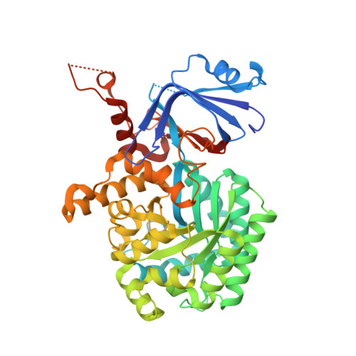
    Guanine deaminase is a metabolic enzyme, found in all forms of life, which catalyzes the conversion of guanine to xanthine. Despite the availability of several crystal structures, the molecular determinants of substrate orientation and mechanism remain to be elucidated for the amidohydrolase family of guanine deaminase enzymes. Here, we report the crystal structures of Escherichia coli and Saccharomyces cerevisiae guanine deaminase enzymes (EcGuaD and Gud1, respectively), both members of the amidohydrolase superfamily. EcGuaD and Gud1 retain the overall TIM barrel tertiary structure conserved among amidohydrolase enzymes. Both proteins also possess a single zinc cation with trigonal bipyrimidal coordination geometry within their active sites. We also determined a liganded structure of Gud1 bound to the product, xanthine. Analysis of this structure, along with kinetic data of native and site-directed mutants of EcGuaD, identifies several key residues that are responsible for substrate recognition and catalysis. In addition, after a small library of compounds had been screened, two guanine derivatives, 8-azaguanine and 1-methylguanine, were identified as EcGuaD substrates. Interestingly, both EcGuaD and Gud1 also exhibit secondary ammeline deaminase activity. Overall, this work details key structural features of substrate recognition and catalysis of the amidohydrolase family of guanine deaminase enzymes in support of our long-term goal to engineer these enzymes with altered activity and substrate specificity.


  • Organizational Affiliation
    • Department of Biochemistry and Cell Biology , Stony Brook University , Stony Brook , New York 11794 , United States.

Macromolecule Content 

  • Total Structure Weight: 57.38 kDa 
  • Atom Count: 4,083 
  • Modeled Residue Count: 452 
  • Deposited Residue Count: 489 
  • Unique protein chains: 1

Macromolecules

Find similar proteins by:|  3D Structure
Entity ID: 1
MoleculeChains  Sequence LengthOrganismDetailsImage
Yeast Guanine Deaminase489Saccharomyces cerevisiae S288CMutation(s): 0 
Gene Names: GUD1YDL238C
EC: 3.5.4.3
UniProt
Find proteins for Q07729 (Saccharomyces cerevisiae (strain ATCC 204508 / S288c))
Explore Q07729 
Go to UniProtKB:  Q07729
Entity Groups
Sequence Clusters30% Identity50% Identity70% Identity90% Identity95% Identity100% Identity
UniProt GroupQ07729
Sequence Annotations
Expand
Reference Sequence

Small Molecules

Ligands 3 Unique
IDChains Name / Formula / InChI Key2D Diagram3D Interactions
2PE

Query on 2PE


Download:Ideal Coordinates CCD File
G [auth A],
H [auth A],
I [auth A],
J [auth A]
NONAETHYLENE GLYCOL
C18 H38 O10
YZUUTMGDONTGTN-UHFFFAOYSA-N
SO4

Query on SO4


Download:Ideal Coordinates CCD File
C [auth A],
D [auth A],
E [auth A],
F [auth A]
SULFATE ION
O4 S
QAOWNCQODCNURD-UHFFFAOYSA-L
ZN

Query on ZN


Download:Ideal Coordinates CCD File
B [auth A]ZINC ION
Zn
PTFCDOFLOPIGGS-UHFFFAOYSA-N

Experimental Data & Validation

Experimental Data

  • Method: X-RAY DIFFRACTION
  • Resolution: 1.75 Å
  • R-Value Free:  0.156 (Depositor), 0.159 (DCC) 
  • R-Value Work:  0.141 (Depositor), 0.144 (DCC) 
  • R-Value Observed: 0.142 (Depositor) 
Space Group: P 43 2 2
Unit Cell:
Length ( Å )Angle ( ˚ )
a = 107.229α = 90
b = 107.229β = 90
c = 114.831γ = 90
Software Package:
Software NamePurpose
PHENIXrefinement
XDSdata reduction
Aimlessdata scaling
PHASERphasing

Structure Validation

View Full Validation Report


Entry History 

& Funding Information

Deposition Data


Funding OrganizationLocationGrant Number
National Institutes of Health/National Institute of General Medical Sciences (NIH/NIGMS)United StatesR35GM124898
National Institutes of Health/National Institute of General Medical Sciences (NIH/NIGMS)United StatesP41GM103403

Revision History  (Full details and data files)

  • Version 1.0: 2019-07-24
    Type: Initial release
  • Version 1.1: 2019-08-14
    Changes: Data collection, Database references
  • Version 1.2: 2020-01-01
    Changes: Author supporting evidence
  • Version 1.3: 2023-10-11
    Changes: Data collection, Database references, Refinement description