6OH2 | pdb_00006oh2

X-ray crystal structure of the mouse CMP-sialic acid transporter in complex with CMP, by lipidic cubic phase


Experimental Data Snapshot

  • Method: X-RAY DIFFRACTION
  • Resolution: 2.58 Å
  • R-Value Free: 
    0.252 (Depositor), 0.263 (DCC) 
  • R-Value Work: 
    0.241 (Depositor), 0.249 (DCC) 
  • R-Value Observed: 
    0.241 (Depositor) 

wwPDB Validation 3D Report Full Report

Validation slider image for 6OH2

Ligand Structure Quality Assessment 


This is version 1.2 of the entry. See complete history

Literature

Structural basis for mammalian nucleotide sugar transport.

Ahuja, S.Whorton, M.R.

(2019) Elife 8

  • DOI: https://doi.org/10.7554/eLife.45221
  • Primary Citation Related Structures: 
    6OH2, 6OH3, 6OH4

  • PubMed Abstract: 

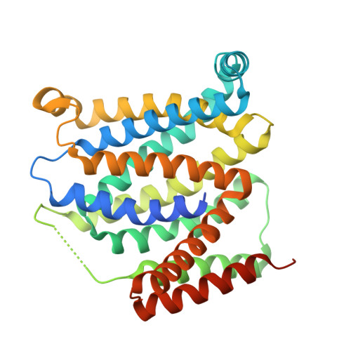
    Nucleotide-sugar transporters (NSTs) are critical components of the cellular glycosylation machinery. They transport nucleotide-sugar conjugates into the Golgi lumen, where they are used for the glycosylation of proteins and lipids, and they then subsequently transport the nucleotide monophosphate byproduct back to the cytoplasm. Dysregulation of human NSTs causes several debilitating diseases, and NSTs are virulence factors for many pathogens. Here we present the first crystal structures of a mammalian NST, the mouse CMP-sialic acid transporter (mCST), in complex with its physiological substrates CMP and CMP-sialic acid. Detailed visualization of extensive protein-substrate interactions explains the mechanisms governing substrate selectivity. Further structural analysis of mCST's unique lumen-facing partially-occluded conformation, coupled with the characterization of substrate-induced quenching of mCST's intrinsic tryptophan fluorescence, reveals the concerted conformational transitions that occur during substrate transport. These results provide a framework for understanding the effects of disease-causing mutations and the mechanisms of this diverse family of transporters.


  • Organizational Affiliation
    • Vollum Institute, Oregon Health & Science University, Portland, United States.

Macromolecule Content 

  • Total Structure Weight: 36.18 kDa 
  • Atom Count: 2,267 
  • Modeled Residue Count: 299 
  • Deposited Residue Count: 330 
  • Unique protein chains: 1

Macromolecules

Find similar proteins by:|  3D Structure
Entity ID: 1
MoleculeChains  Sequence LengthOrganismDetailsImage
CMP-sialic acid transporter330Mus musculusMutation(s): 0 
Gene Names: Slc35a1
Membrane Entity: Yes 
UniProt
Find proteins for Q61420 (Mus musculus)
Explore Q61420 
Go to UniProtKB:  Q61420
Entity Groups
Sequence Clusters30% Identity50% Identity70% Identity90% Identity95% Identity100% Identity
UniProt GroupQ61420
Sequence Annotations
Expand
Reference Sequence

Small Molecules

Ligands 1 Unique
IDChains Name / Formula / InChI Key2D Diagram3D Interactions
C5P
(Subject of Investigation/LOI)

Query on C5P


Download:Ideal Coordinates CCD File
B [auth A]CYTIDINE-5'-MONOPHOSPHATE
C9 H14 N3 O8 P
IERHLVCPSMICTF-XVFCMESISA-N

Experimental Data & Validation

Experimental Data

  • Method: X-RAY DIFFRACTION
  • Resolution: 2.58 Å
  • R-Value Free:  0.252 (Depositor), 0.263 (DCC) 
  • R-Value Work:  0.241 (Depositor), 0.249 (DCC) 
  • R-Value Observed: 0.241 (Depositor) 
Space Group: C 1 2 1
Unit Cell:
Length ( Å )Angle ( ˚ )
a = 144.54α = 90
b = 49.53β = 107.63
c = 50.15γ = 90
Software Package:
Software NamePurpose
REFMACrefinement
XDSdata reduction
XDSdata scaling
PHASERphasing

Structure Validation

View Full Validation Report


Ligand Structure Quality Assessment 


Entry History 

& Funding Information

Deposition Data


Funding OrganizationLocationGrant Number
National Institutes of Health/National Institute of General Medical Sciences (NIH/NIGMS)United StatesR01GM130909

Revision History  (Full details and data files)

  • Version 1.0: 2019-04-24
    Type: Initial release
  • Version 1.1: 2020-01-01
    Changes: Author supporting evidence
  • Version 1.2: 2024-03-13
    Changes: Data collection, Database references