6OE9 | pdb_00006oe9

Crystal structure of p204 HIN1 domain


Experimental Data Snapshot

  • Method: X-RAY DIFFRACTION
  • Resolution: 1.94 Å
  • R-Value Free: 
    0.192 (Depositor), 0.201 (DCC) 
  • R-Value Work: 
    0.172 (Depositor), 0.182 (DCC) 
  • R-Value Observed: 
    0.174 (Depositor) 

Starting Model: experimental
View more details

wwPDB Validation 3D Report Full Report

Validation slider image for 6OE9

This is version 1.2 of the entry. See complete history

Literature

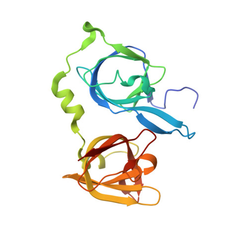
Structural analysis of the HIN1 domain of interferon-inducible protein 204.

Tian, Y.Yin, Q.

(2019) Acta Crystallogr F Struct Biol Commun 75: 455-460

  • DOI: https://doi.org/10.1107/S2053230X19007167
  • Primary Citation Related Structures: 
    6OE9

  • PubMed Abstract: 

    Interferon-inducible protein 204 (p204) binds to microbial DNA to elicit inflammatory responses and induce interferon production. p204 also modulates cell proliferation and differentiation by regulating various transcription factors. The C-terminal HIN domains in p204 are believed to be responsible for DNA binding, but the binding mode is not fully understood. The DNA-binding affinity of the p204 HIN1 domain has been characterized and its crystal structure has been determined, providing insight into its interaction with DNA. Surface-charge distribution together with sequence alignment suggests that the p204 HIN domain uses its L12 and L45 loops for DNA binding.


  • Organizational Affiliation
    • Department of Biological Science, Florida State University, Tallahassee, FL 32306, USA.

Macromolecule Content 

  • Total Structure Weight: 23.89 kDa 
  • Atom Count: 1,843 
  • Modeled Residue Count: 201 
  • Deposited Residue Count: 205 
  • Unique protein chains: 1

Macromolecules

Find similar proteins by:|  3D Structure
Entity ID: 1
MoleculeChains  Sequence LengthOrganismDetailsImage
Interferon-activable protein 204205Mus musculusMutation(s): 0 
Gene Names: Ifi204
UniProt & NIH Common Fund Data Resources
Find proteins for P0DOV2 (Mus musculus)
Explore P0DOV2 
Go to UniProtKB:  P0DOV2
IMPC:  MGI:96429
Entity Groups
Sequence Clusters30% Identity50% Identity70% Identity90% Identity95% Identity100% Identity
UniProt GroupP0DOV2
Sequence Annotations
Expand
Reference Sequence

Small Molecules

Ligands 2 Unique
IDChains Name / Formula / InChI Key2D Diagram3D Interactions
SO4

Query on SO4


Download:Ideal Coordinates CCD File
F [auth A],
G [auth A],
H [auth A],
I [auth A]
SULFATE ION
O4 S
QAOWNCQODCNURD-UHFFFAOYSA-L
GOL

Query on GOL


Download:Ideal Coordinates CCD File
B [auth A],
C [auth A],
D [auth A],
E [auth A]
GLYCEROL
C3 H8 O3
PEDCQBHIVMGVHV-UHFFFAOYSA-N

Experimental Data & Validation

Experimental Data

  • Method: X-RAY DIFFRACTION
  • Resolution: 1.94 Å
  • R-Value Free:  0.192 (Depositor), 0.201 (DCC) 
  • R-Value Work:  0.172 (Depositor), 0.182 (DCC) 
  • R-Value Observed: 0.174 (Depositor) 
Space Group: P 65 2 2
Unit Cell:
Length ( Å )Angle ( ˚ )
a = 58.383α = 90
b = 58.383β = 90
c = 315.836γ = 120
Software Package:
Software NamePurpose
PHENIXrefinement
HKL-2000data reduction
HKL-2000data scaling
PHASERphasing

Structure Validation

View Full Validation Report


Entry History 

& Funding Information

Deposition Data


Funding OrganizationLocationGrant Number
National Institutes of Health/National Human Genome Research Institute (NIH/NHGRI)United StatesR00AI108793

Revision History  (Full details and data files)

  • Version 1.0: 2019-07-10
    Type: Initial release
  • Version 1.1: 2019-12-18
    Changes: Author supporting evidence
  • Version 1.2: 2023-10-11
    Changes: Data collection, Database references, Refinement description