6OC1 | pdb_00006oc1

Crystal structure of human DHODH with TAK-632


Experimental Data Snapshot

  • Method: X-RAY DIFFRACTION
  • Resolution: 2.70 Å
  • R-Value Free: 
    0.294 (Depositor), 0.291 (DCC) 
  • R-Value Work: 
    0.230 (Depositor), 0.234 (DCC) 
  • R-Value Observed: 
    0.233 (Depositor) 

Starting Model: experimental
View more details

wwPDB Validation 3D Report Full Report

Validation slider image for 6OC1

Ligand Structure Quality Assessment 


This is version 1.3 of the entry. See complete history

Literature

Metabolic Modifier Screen Reveals Secondary Targets of Protein Kinase Inhibitors within Nucleotide Metabolism.

Abt, E.R.Rosser, E.W.Durst, M.A.Lok, V.Poddar, S.Le, T.M.Cho, A.Kim, W.Wei, L.Song, J.Capri, J.R.Xu, S.Wu, N.Slavik, R.Jung, M.E.Damoiseaux, R.Czernin, J.Donahue, T.R.Lavie, A.Radu, C.G.

(2020) Cell Chem Biol 27: 197

  • DOI: https://doi.org/10.1016/j.chembiol.2019.10.012
  • Primary Citation Related Structures: 
    6OC0, 6OC1

  • PubMed Abstract: 

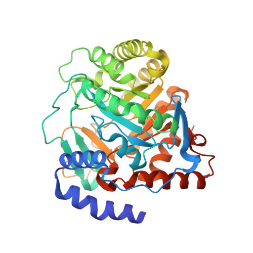
    Biosynthesis of the pyrimidine nucleotide uridine monophosphate (UMP) is essential for cell proliferation and is achieved by the activity of convergent de novo and salvage metabolic pathways. Here we report the development and application of a cell-based metabolic modifier screening platform that leverages the redundancy in pyrimidine metabolism for the discovery of selective UMP biosynthesis modulators. In evaluating a library of protein kinase inhibitors, we identified multiple compounds that possess nucleotide metabolism modifying activity. The JNK inhibitor JNK-IN-8 was found to potently inhibit nucleoside transport and engage ENT1. The PDK1 inhibitor OSU-03012 (also known as AR-12) and the RAF inhibitor TAK-632 were shown to inhibit the therapeutically relevant de novo pathway enzyme DHODH and their affinities were unambiguously confirmed through in vitro assays and co-crystallization with human DHODH.


  • Organizational Affiliation
    • Department of Molecular and Medical Pharmacology, University of California Los Angeles, Los Angeles, CA, USA; Ahmanson Translational Imaging Division, University of California Los Angeles, Los Angeles, CA, USA.

Macromolecule Content 

  • Total Structure Weight: 41.39 kDa 
  • Atom Count: 2,891 
  • Modeled Residue Count: 363 
  • Deposited Residue Count: 369 
  • Unique protein chains: 1

Macromolecules

Find similar proteins by:|  3D Structure
Entity ID: 1
MoleculeChains  Sequence LengthOrganismDetailsImage
Dihydroorotate dehydrogenase (quinone), mitochondrial369Homo sapiensMutation(s): 0 
Gene Names: DHODH
EC: 1.3.5.2
UniProt & NIH Common Fund Data Resources
Find proteins for Q02127 (Homo sapiens)
Explore Q02127 
Go to UniProtKB:  Q02127
PHAROS:  Q02127
GTEx:  ENSG00000102967 
Entity Groups
Sequence Clusters30% Identity50% Identity70% Identity90% Identity95% Identity100% Identity
UniProt GroupQ02127
Sequence Annotations
Expand
Reference Sequence

Small Molecules

Experimental Data & Validation

Experimental Data

  • Method: X-RAY DIFFRACTION
  • Resolution: 2.70 Å
  • R-Value Free:  0.294 (Depositor), 0.291 (DCC) 
  • R-Value Work:  0.230 (Depositor), 0.234 (DCC) 
  • R-Value Observed: 0.233 (Depositor) 
Space Group: P 21 3
Unit Cell:
Length ( Å )Angle ( ˚ )
a = 113.668α = 90
b = 113.668β = 90
c = 113.668γ = 90
Software Package:
Software NamePurpose
REFMACrefinement
MOLREPphasing
XDSdata reduction
Cootmodel building
XDSdata scaling

Structure Validation

View Full Validation Report


Ligand Structure Quality Assessment 


Entry History 

Deposition Data

Revision History  (Full details and data files)

  • Version 1.0: 2019-11-13
    Type: Initial release
  • Version 1.1: 2019-11-27
    Changes: Database references
  • Version 1.2: 2020-03-04
    Changes: Database references
  • Version 1.3: 2023-10-11
    Changes: Data collection, Database references, Refinement description