6OAY | pdb_00006oay

Structure of the hyperactive ClpB mutant K476C, bound to casein, post-state


Experimental Data Snapshot

  • Method: ELECTRON MICROSCOPY
  • Resolution: 3.30 Å
  • Aggregation State: PARTICLE 
  • Reconstruction Method: SINGLE PARTICLE 

wwPDB Validation 3D Report Full Report

Validation slider image for 6OAY

This is version 1.3 of the entry. See complete history

Literature

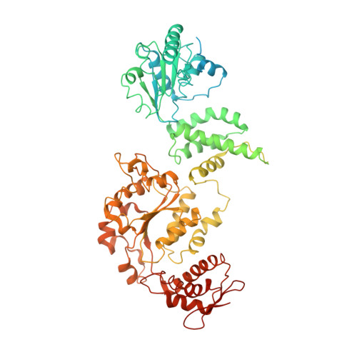
Structural basis for substrate gripping and translocation by the ClpB AAA+ disaggregase.

Rizo, A.N.Lin, J.Gates, S.N.Tse, E.Bart, S.M.Castellano, L.M.DiMaio, F.Shorter, J.Southworth, D.R.

(2019) Nat Commun 10: 2393-2393

  • DOI: https://doi.org/10.1038/s41467-019-10150-y
  • Primary Citation Related Structures: 
    6OAX, 6OAY, 6OG1, 6OG2, 6OG3

  • PubMed Abstract: 

    Bacterial ClpB and yeast Hsp104 are homologous Hsp100 protein disaggregases that serve critical functions in proteostasis by solubilizing protein aggregates. Two AAA+ nucleotide binding domains (NBDs) power polypeptide translocation through a central channel comprised of a hexameric spiral of protomers that contact substrate via conserved pore-loop interactions. Here we report cryo-EM structures of a hyperactive ClpB variant bound to the model substrate, casein in the presence of slowly hydrolysable ATPγS, which reveal the translocation mechanism. Distinct substrate-gripping interactions are identified for NBD1 and NBD2 pore loops. A trimer of N-terminal domains define a channel entrance that binds the polypeptide substrate adjacent to the topmost NBD1 contact. NBD conformations at the seam interface reveal how ATP hydrolysis-driven substrate disengagement and re-binding are precisely tuned to drive a directional, stepwise translocation cycle.


  • Organizational Affiliation
    • Graduate Program in Chemical Biology, University of Michigan, Ann Arbor, MI, 48109, USA.

Macromolecule Content 

  • Total Structure Weight: 589.71 kDa 
  • Atom Count: 27,261 
  • Modeled Residue Count: 3,412 
  • Deposited Residue Count: 5,228 
  • Unique protein chains: 2

Macromolecules

Find similar proteins by:|  3D Structure
Entity ID: 1
MoleculeChains  Sequence LengthOrganismDetailsImage
Hyperactive disaggregase ClpB867Escherichia coli K-12Mutation(s): 0 
UniProt
Find proteins for P63284 (Escherichia coli (strain K12))
Explore P63284 
Go to UniProtKB:  P63284
Entity Groups
Sequence Clusters30% Identity50% Identity70% Identity90% Identity95% Identity100% Identity
UniProt GroupP63284
Sequence Annotations
Expand
Reference Sequence
Find similar proteins by:|  3D Structure
Entity ID: 2
MoleculeChains  Sequence LengthOrganismDetailsImage
Alpha-S1-caseinG [auth P]26Bos taurusMutation(s): 0 

Small Molecules

Ligands 2 Unique
IDChains Name / Formula / InChI Key2D Diagram3D Interactions
AGS

Query on AGS


Download:Ideal Coordinates CCD File
H [auth C]
I [auth C]
K [auth E]
L [auth E]
M [auth D]
H [auth C],
I [auth C],
K [auth E],
L [auth E],
M [auth D],
N [auth D],
O [auth B]
PHOSPHOTHIOPHOSPHORIC ACID-ADENYLATE ESTER
C10 H16 N5 O12 P3 S
NLTUCYMLOPLUHL-KQYNXXCUSA-N
ADP

Query on ADP


Download:Ideal Coordinates CCD File
J [auth F],
P [auth B],
Q [auth A],
R [auth A]
ADENOSINE-5'-DIPHOSPHATE
C10 H15 N5 O10 P2
XTWYTFMLZFPYCI-KQYNXXCUSA-N

Experimental Data & Validation

Experimental Data

  • Method: ELECTRON MICROSCOPY
  • Resolution: 3.30 Å
  • Aggregation State: PARTICLE 
  • Reconstruction Method: SINGLE PARTICLE 
EM Software:
TaskSoftware PackageVersion
MODEL REFINEMENTPHENIX

Structure Validation

View Full Validation Report


Entry History 

Deposition Data

Revision History  (Full details and data files)

  • Version 1.0: 2019-06-12
    Type: Initial release
  • Version 1.1: 2019-06-19
    Changes: Data collection, Database references
  • Version 1.2: 2024-03-20
    Changes: Data collection, Database references, Derived calculations
  • Version 1.3: 2025-05-14
    Changes: Data collection, Structure summary