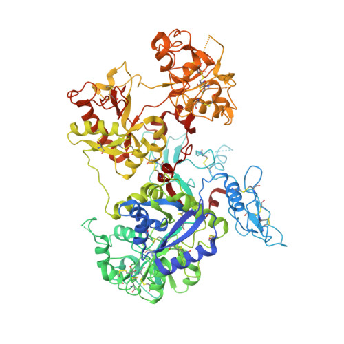
6O0D

Saxiphilin Apo structure

  • Classification: ANTITOXIN
  • Organism(s): Aquarana catesbeiana
  • Expression System: Spodoptera frugiperda
  • Mutation(s): No 

  • Deposited: 2019-02-16 Released: 2019-07-10 
  • Deposition Author(s): Yen, T.J., Lolicato, M., Minor, D.L.
  • Funding Organization(s): National Institutes of Health/National Heart, Lung, and Blood Institute (NIH/NHLBI), National Institutes of Health/National Institute of General Medical Sciences (NIH/NIGMS)

Experimental Data Snapshot

  • Method: X-RAY DIFFRACTION
  • Resolution: 2.50 Å
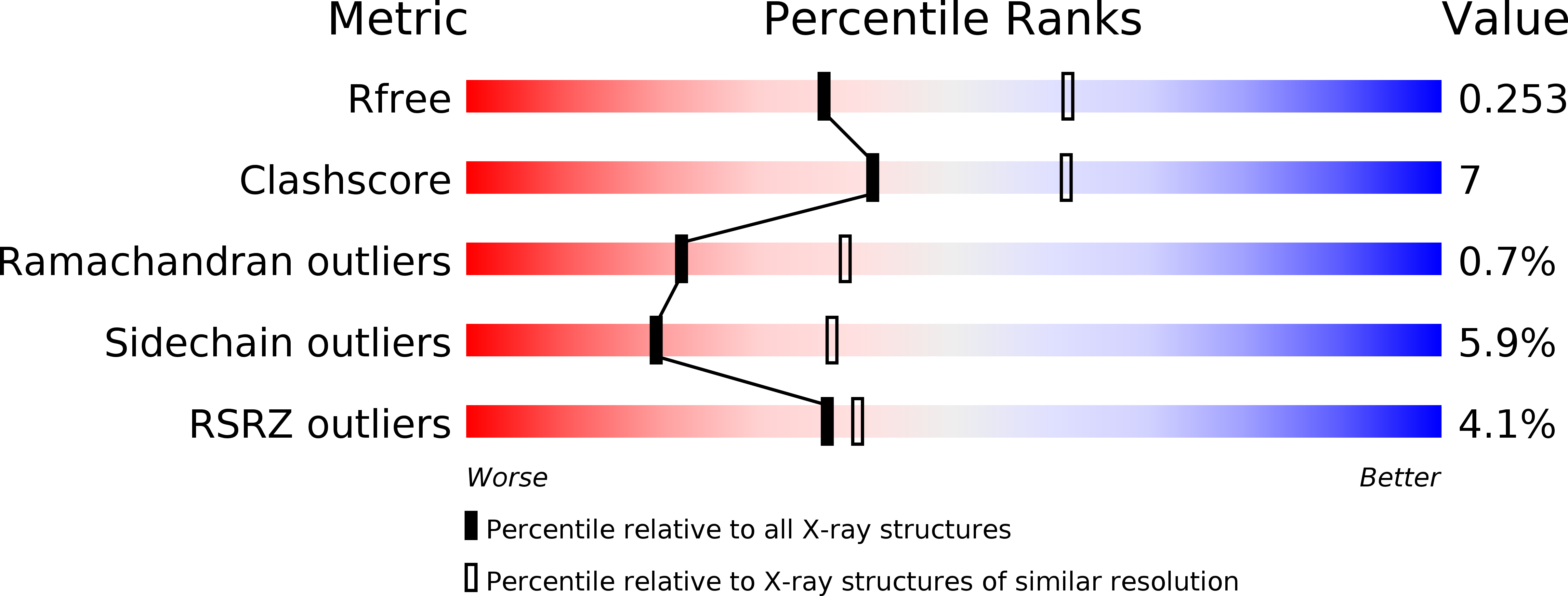
  • R-Value Free: 0.253 
  • R-Value Work: 0.224 
  • R-Value Observed: 0.226 

wwPDB Validation   3D Report Full Report


This is version 1.1 of the entry. See complete history


Literature

Structure of the saxiphilin:saxitoxin (STX) complex reveals a convergent molecular recognition strategy for paralytic toxins.

Yen, T.J.Lolicato, M.Thomas-Tran, R.Du Bois, J.Minor Jr., D.L.

(2019) Sci Adv 5: eaax2650-eaax2650

  • DOI: https://doi.org/10.1126/sciadv.aax2650
  • Primary Citation of Related Structures:  
    6O0D, 6O0E, 6O0F

  • PubMed Abstract: 

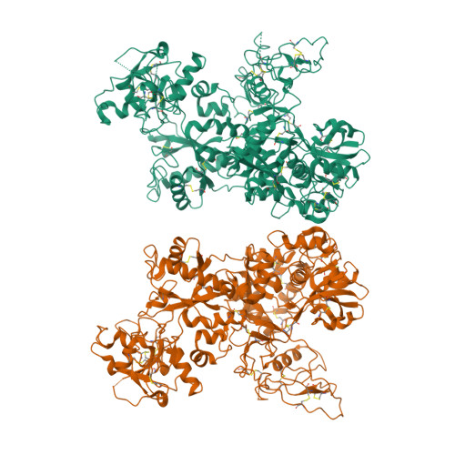
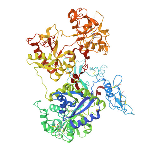
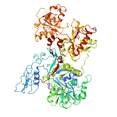
    Dinoflagelates and cyanobacteria produce saxitoxin (STX), a lethal bis-guanidinium neurotoxin causing paralytic shellfish poisoning. A number of metazoans have soluble STX-binding proteins that may prevent STX intoxication. However, their STX molecular recognition mechanisms remain unknown. Here, we present structures of saxiphilin (Sxph), a bullfrog high-affinity STX-binding protein, alone and bound to STX. The structures reveal a novel high-affinity STX-binding site built from a "proto-pocket" on a transferrin scaffold that also bears thyroglobulin domain protease inhibitor repeats. Comparison of Sxph and voltage-gated sodium channel STX-binding sites reveals a convergent toxin recognition strategy comprising a largely rigid binding site where acidic side chains and a cation-π interaction engage STX. These studies reveal molecular rules for STX recognition, outline how a toxin-binding site can be built on a naïve scaffold, and open a path to developing protein sensors for environmental STX monitoring and new biologics for STX intoxication mitigation.


  • Organizational Affiliation

    Cardiovascular Research Institute, University of California, San Francisco, San Francisco, CA 94158, USA.


Macromolecules
Find similar proteins by:  (by identity cutoff)  |  3D Structure
Entity ID: 1
MoleculeChains Sequence LengthOrganismDetailsImage
Saxiphilin
A, B
853Aquarana catesbeianaMutation(s): 0 
UniProt
Find proteins for P31226 (Aquarana catesbeiana)
Explore P31226 
Go to UniProtKB:  P31226
Entity Groups  
Sequence Clusters30% Identity50% Identity70% Identity90% Identity95% Identity100% Identity
UniProt GroupP31226
Sequence Annotations
Expand
  • Reference Sequence
Experimental Data & Validation

Experimental Data

  • Method: X-RAY DIFFRACTION
  • Resolution: 2.50 Å
  • R-Value Free: 0.253 
  • R-Value Work: 0.224 
  • R-Value Observed: 0.226 
  • Space Group: P 21 21 21
Unit Cell:
Length ( Å )Angle ( ˚ )
a = 96.158α = 90
b = 111.309β = 90
c = 254.834γ = 90
Software Package:
Software NamePurpose
PHENIXrefinement
Aimlessdata scaling
XDSdata reduction
PHASERphasing

Structure Validation

View Full Validation Report


Entry History & Funding Information

Deposition Data


Funding OrganizationLocationGrant Number
National Institutes of Health/National Heart, Lung, and Blood Institute (NIH/NHLBI)United StatesR01-HL080050
National Institutes of Health/National Institute of General Medical Sciences (NIH/NIGMS)United StatesGM117263-01A1

Revision History  (Full details and data files)

  • Version 1.0: 2019-07-10
    Type: Initial release
  • Version 1.1: 2019-12-04
    Changes: Author supporting evidence