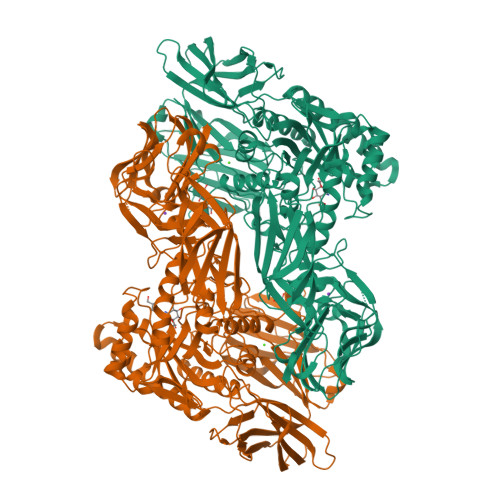
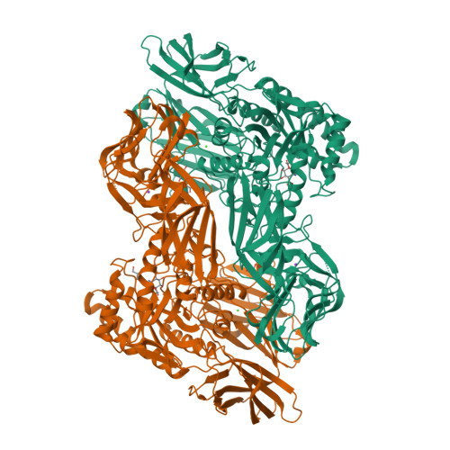
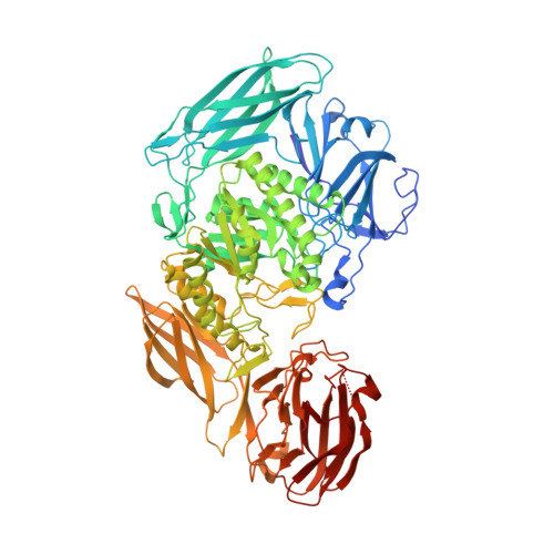
6NZG

Bacteroides uniformis beta-glucuronidase 2 covalently bound to cyclophellitol-6-carboxylate aziridine


Experimental Data Snapshot

  • Method: X-RAY DIFFRACTION
  • Resolution: 2.43 Å
  • R-Value Free: 0.221 
  • R-Value Work: 0.170 
  • R-Value Observed: 0.172 

Starting Model: experimental
View more details

wwPDB Validation   3D Report Full Report


Ligand Structure Quality Assessment 


This is version 2.2 of the entry. See complete history


Literature

Discovering the Microbial Enzymes Driving Drug Toxicity with Activity-Based Protein Profiling.

Jariwala, P.B.Pellock, S.J.Goldfarb, D.Cloer, E.W.Artola, M.Simpson, J.B.Bhatt, A.P.Walton, W.G.Roberts, L.R.Major, M.B.Davies, G.J.Overkleeft, H.S.Redinbo, M.R.

(2020) ACS Chem Biol 15: 217-225

  • DOI: https://doi.org/10.1021/acschembio.9b00788
  • Primary Citation of Related Structures:  
    6NZG

  • PubMed Abstract: 

    It is increasingly clear that interindividual variability in human gut microbial composition contributes to differential drug responses. For example, gastrointestinal (GI) toxicity is not observed in all patients treated with the anticancer drug irinotecan, and it has been suggested that this variability is a result of differences in the types and levels of gut bacterial β-glucuronidases (GUSs). GUS enzymes promote drug toxicity by hydrolyzing the inactive drug-glucuronide conjugate back to the active drug, which damages the GI epithelium. Proteomics-based identification of the exact GUS enzymes responsible for drug reactivation from the complexity of the human microbiota has not been accomplished, however. Here, we discover the specific bacterial GUS enzymes that generate SN-38, the active and toxic metabolite of irinotecan, from human fecal samples using a unique activity-based protein profiling (ABPP) platform. We identify and quantify gut bacterial GUS enzymes from human feces with an ABPP-enabled proteomics pipeline and then integrate this information with ex vivo kinetics to pinpoint the specific GUS enzymes responsible for SN-38 reactivation. Furthermore, the same approach also reveals the molecular basis for differential gut bacterial GUS inhibition observed between human fecal samples. Taken together, this work provides an unprecedented technical and bioinformatics pipeline to discover the microbial enzymes responsible for specific reactions from the complexity of human feces. Identifying such microbial enzymes may lead to precision biomarkers and novel drug targets to advance the promise of personalized medicine.


  • Organizational Affiliation

    Department of Bioorganic Synthesis, Leiden Institute of Chemistry , Leiden University , Leiden 2311 , The Netherlands.


Macromolecules
Find similar proteins by:  (by identity cutoff)  |  3D Structure
Entity ID: 1
MoleculeChains Sequence LengthOrganismDetailsImage
Beta-galactosidase
A, B
878Bacteroides uniformisMutation(s): 0 
Gene Names: uidA_3ERS417307_01034
EC: 3.2.1.23 (PDB Primary Data), 3.2.1.31 (PDB Primary Data)
UniProt
Find proteins for A0A414F142 (Bacteroides uniformis)
Explore A0A414F142 
Go to UniProtKB:  A0A414F142
Entity Groups  
Sequence Clusters30% Identity50% Identity70% Identity90% Identity95% Identity100% Identity
UniProt GroupA0A414F142
Sequence Annotations
Expand
  • Reference Sequence
Small Molecules
Ligands 4 Unique
IDChains Name / Formula / InChI Key2D Diagram3D Interactions
L9D
Query on L9D

Download Ideal Coordinates CCD File 
C [auth A],
G [auth B]
(1S,2R,3S,4S,5S,6R)-2-amino-3,4,5,6-tetrahydroxycyclohexane-1-carboxylic acid
C7 H13 N O6
POIWYXZEYSYIEW-HOZKJCLWSA-N
GOL
Query on GOL

Download Ideal Coordinates CCD File 
D [auth A],
H [auth B],
I [auth B],
J [auth B]
GLYCEROL
C3 H8 O3
PEDCQBHIVMGVHV-UHFFFAOYSA-N
CA
Query on CA

Download Ideal Coordinates CCD File 
F [auth A],
L [auth B]
CALCIUM ION
Ca
BHPQYMZQTOCNFJ-UHFFFAOYSA-N
K
Query on K

Download Ideal Coordinates CCD File 
E [auth A],
K [auth B]
POTASSIUM ION
K
NPYPAHLBTDXSSS-UHFFFAOYSA-N
Experimental Data & Validation

Experimental Data

  • Method: X-RAY DIFFRACTION
  • Resolution: 2.43 Å
  • R-Value Free: 0.221 
  • R-Value Work: 0.170 
  • R-Value Observed: 0.172 
  • Space Group: P 21 21 21
Unit Cell:
Length ( Å )Angle ( ˚ )
a = 74.536α = 90
b = 142.407β = 90
c = 180.664γ = 90
Software Package:
Software NamePurpose
PHENIXrefinement
XDSdata reduction
Aimlessdata scaling
PHENIXphasing

Structure Validation

View Full Validation Report


Ligand Structure Quality Assessment 


Entry History & Funding Information

Deposition Data


Funding OrganizationLocationGrant Number
National Institutes of Health/National Cancer Institute (NIH/NCI)United States--

Revision History  (Full details and data files)

  • Version 1.0: 2019-12-18
    Type: Initial release
  • Version 2.0: 2019-12-25
    Changes: Advisory, Derived calculations, Non-polymer description, Structure summary
  • Version 2.1: 2020-01-29
    Changes: Database references
  • Version 2.2: 2023-10-11
    Changes: Data collection, Database references, Derived calculations, Refinement description