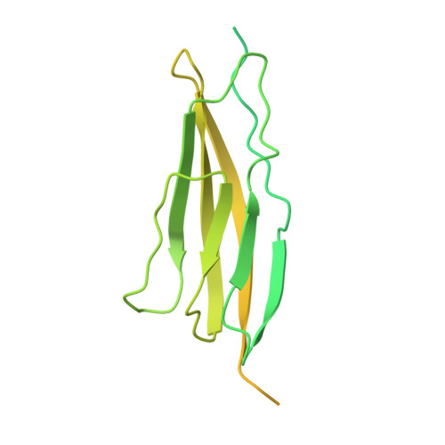
RCSB PDB - 6NZ0: Cryo-EM structure of AAV-2 in complex with AAVR PKD domains 1 and 2

 6NZ0

Cryo-EM structure of AAV-2 in complex with AAVR PKD domains 1 and 2


Experimental Data Snapshot

  • Method: ELECTRON MICROSCOPY
  • Resolution: 2.40 Å
  • Aggregation State: PARTICLE 
  • Reconstruction Method: SINGLE PARTICLE 

Starting Model: experimental
View more details

wwPDB Validation   3D Report Full Report


This is version 1.3 of the entry. See complete history


Literature

Structure of the gene therapy vector, adeno-associated virus with its cell receptor, AAVR.

Meyer, N.L.Hu, G.Davulcu, O.Xie, Q.Noble, A.J.Yoshioka, C.Gingerich, D.S.Trzynka, A.David, L.Stagg, S.M.Chapman, M.S.

(2019) Elife 8

  • DOI: https://doi.org/10.7554/eLife.44707
  • Primary Citation of Related Structures:  
    6NZ0

  • PubMed Abstract: 

    Adeno-associated virus (AAV) vectors are preeminent in emerging clinical gene therapies. Generalizing beyond the most tractable genetic diseases will require modulation of cell specificity and immune neutralization. Interactions of AAV with its cellular receptor, AAVR, are key to understanding cell-entry and trafficking with the rigor needed to engineer tissue-specific vectors. Cryo -electron tomography shows ordered binding of part of the flexible receptor to the viral surface, with distal domains in multiple conformations. Regions of the virus and receptor in close physical proximity can be identified by cross-linking/mass spectrometry. Cryo -electron microscopy with a two-domain receptor fragment reveals the interactions at 2.4 Å resolution. AAVR binds between AAV's spikes on a plateau that is conserved, except in one clade whose structure is AAVR-incompatible. AAVR's footprint overlaps the epitopes of several neutralizing antibodies, prompting a re-evaluation of neutralization mechanisms. The structure provides a roadmap for experimental probing and manipulation of viral-receptor interactions.


  • Organizational Affiliation

    Department of Biochemistry and Molecular Biology, Oregon Health and Science University, Portland, United States.


Macromolecules
Find similar proteins by:  (by identity cutoff)  |  3D Structure
Entity ID: 1
MoleculeChains Sequence LengthOrganismDetailsImage
Dyslexia-associated protein KIAA0319-like proteinA [auth Z]290Homo sapiensMutation(s): 0 
Gene Names: KIAA0319LAAVRKIAA1837PP791
UniProt & NIH Common Fund Data Resources
Find proteins for Q8IZA0 (Homo sapiens)
Explore Q8IZA0 
Go to UniProtKB:  Q8IZA0
PHAROS:  Q8IZA0
GTEx:  ENSG00000142687 
Entity Groups  
Sequence Clusters30% Identity50% Identity70% Identity90% Identity95% Identity100% Identity
UniProt GroupQ8IZA0
Sequence Annotations
Expand
  • Reference Sequence
Find similar proteins by:  (by identity cutoff)  |  3D Structure
Entity ID: 2
MoleculeChains Sequence LengthOrganismDetailsImage
Capsid protein VP1B [auth A]735Adeno-associated virus 2 Srivastava/1982Mutation(s): 0 
Gene Names: VP1
UniProt
Find proteins for P03135 (Adeno-associated virus 2 (isolate Srivastava/1982))
Explore P03135 
Go to UniProtKB:  P03135
Entity Groups  
Sequence Clusters30% Identity50% Identity70% Identity90% Identity95% Identity100% Identity
UniProt GroupP03135
Sequence Annotations
Expand
  • Reference Sequence
Small Molecules
Ligands 1 Unique
IDChains Name / Formula / InChI Key2D Diagram3D Interactions
MG
Query on MG

Download Ideal Coordinates CCD File 
C [auth Z]MAGNESIUM ION
Mg
JLVVSXFLKOJNIY-UHFFFAOYSA-N
Experimental Data & Validation

Experimental Data

  • Method: ELECTRON MICROSCOPY
  • Resolution: 2.40 Å
  • Aggregation State: PARTICLE 
  • Reconstruction Method: SINGLE PARTICLE 
EM Software:
TaskSoftware PackageVersion
RECONSTRUCTIONRELION3.0b
MODEL REFINEMENTRSRef

Structure Validation

View Full Validation Report


Entry History & Funding Information

Deposition Data


Funding OrganizationLocationGrant Number
National Institutes of Health/National Institute of General Medical Sciences (NIH/NIGMS)United StatesR35GM122564
National Institutes of Health/National Institute of General Medical Sciences (NIH/NIGMS)United StatesR01GM066875

Revision History  (Full details and data files)

  • Version 1.0: 2019-06-12
    Type: Initial release
  • Version 1.1: 2019-06-19
    Changes: Data collection, Database references
  • Version 1.2: 2019-12-18
    Changes: Author supporting evidence, Other
  • Version 1.3: 2024-10-16
    Changes: Data collection, Database references, Derived calculations, Refinement description, Structure summary