6NYY | pdb_00006nyy

human m-AAA protease AFG3L2, substrate-bound


Experimental Data Snapshot

  • Method: ELECTRON MICROSCOPY
  • Resolution: 3.00 Å
  • Aggregation State: PARTICLE 
  • Reconstruction Method: SINGLE PARTICLE 

Starting Model: experimental
View more details

wwPDB Validation 3D Report Full Report

Validation slider image for 6NYY

This is version 1.5 of the entry. See complete history

Literature

Unique Structural Features of the Mitochondrial AAA+ Protease AFG3L2 Reveal the Molecular Basis for Activity in Health and Disease.

Puchades, C.Ding, B.Song, A.Wiseman, R.L.Lander, G.C.Glynn, S.E.

(2019) Mol Cell 75: 1073

  • DOI: https://doi.org/10.1016/j.molcel.2019.06.016
  • Primary Citation Related Structures: 
    6NYY

  • PubMed Abstract: 

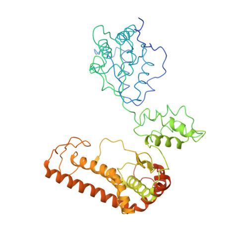
    Mitochondrial AAA+ quality-control proteases regulate diverse aspects of mitochondrial biology through specialized protein degradation, but the underlying mechanisms of these enzymes remain poorly defined. The mitochondrial AAA+ protease AFG3L2 is of particular interest, as genetic mutations localized throughout AFG3L2 are linked to diverse neurodegenerative disorders. However, a lack of structural data has limited our understanding of how mutations impact enzymatic function. Here, we used cryoelectron microscopy (cryo-EM) to determine a substrate-bound structure of the catalytic core of human AFG3L2. This structure identifies multiple specialized structural features that integrate with conserved motifs required for ATP-dependent translocation to unfold and degrade targeted proteins. Many disease-relevant mutations localize to these unique structural features of AFG3L2 and distinctly influence its activity and stability. Our results provide a molecular basis for neurological phenotypes associated with different AFG3L2 mutations and establish a structural framework to understand how different members of the AAA+ superfamily achieve specialized biological functions.


  • Organizational Affiliation
    • Department of Integrative Structural and Computational Biology, The Scripps Research Institute, La Jolla, CA 92037, USA; Department of Molecular Medicine, The Scripps Research Institute, La Jolla, CA 92037, USA; Skaggs Graduate School of Chemical and Biological Sciences, The Scripps Research Institute, La Jolla, CA 92037, USA.

Macromolecule Content 

  • Total Structure Weight: 355.6 kDa 
  • Atom Count: 19,226 
  • Modeled Residue Count: 2,894 
  • Deposited Residue Count: 3,218 
  • Unique protein chains: 2

Macromolecules

Find similar proteins by:|  3D Structure
Entity ID: 1
MoleculeChains  Sequence LengthOrganismDetailsImage
AFG3-like protein 2
A, B, C, D, E
A, B, C, D, E, F
529Homo sapiensMutation(s): 0 
Gene Names: AFG3L2
EC: 3.4.24 (PDB Primary Data), 3.6 (UniProt)
UniProt & NIH Common Fund Data Resources
Find proteins for Q9Y4W6 (Homo sapiens)
Explore Q9Y4W6 
Go to UniProtKB:  Q9Y4W6
PHAROS:  Q9Y4W6
GTEx:  ENSG00000141385 
Entity Groups
Sequence Clusters30% Identity50% Identity70% Identity90% Identity95% Identity100% Identity
UniProt GroupQ9Y4W6
Sequence Annotations
Expand
Reference Sequence
Find similar proteins by:  Sequence   |   3D Structure  
Entity ID: 2
MoleculeChains  Sequence LengthOrganismDetailsImage
Substrate
G, H, I, J
11Homo sapiensMutation(s): 0 

Small Molecules

Ligands 4 Unique
IDChains Name / Formula / InChI Key2D Diagram3D Interactions
ANP

Query on ANP


Download:Ideal Coordinates CCD File
L [auth B],
Q [auth C],
R [auth D]
PHOSPHOAMINOPHOSPHONIC ACID-ADENYLATE ESTER
C10 H17 N6 O12 P3
PVKSNHVPLWYQGJ-KQYNXXCUSA-N
ADP

Query on ADP


Download:Ideal Coordinates CCD File
U [auth E]ADENOSINE-5'-DIPHOSPHATE
C10 H15 N5 O10 P2
XTWYTFMLZFPYCI-KQYNXXCUSA-N
ZN

Query on ZN


Download:Ideal Coordinates CCD File
K [auth A]
N [auth B]
P [auth C]
T [auth D]
V [auth E]
K [auth A],
N [auth B],
P [auth C],
T [auth D],
V [auth E],
W [auth F]
ZINC ION
Zn
PTFCDOFLOPIGGS-UHFFFAOYSA-N
MG

Query on MG


Download:Ideal Coordinates CCD File
M [auth B],
O [auth C],
S [auth D]
MAGNESIUM ION
Mg
JLVVSXFLKOJNIY-UHFFFAOYSA-N

Experimental Data & Validation

Experimental Data

  • Method: ELECTRON MICROSCOPY
  • Resolution: 3.00 Å
  • Aggregation State: PARTICLE 
  • Reconstruction Method: SINGLE PARTICLE 
EM Software:
TaskSoftware PackageVersion
RECONSTRUCTIONRELION3.0
MODEL REFINEMENTPHENIX1.11.1_2580

Structure Validation

View Full Validation Report


Entry History 

& Funding Information

Deposition Data


Funding OrganizationLocationGrant Number
National Institutes of Health/National Cancer Institute (NIH/NCI)United StatesR21 AG061697 01

Revision History  (Full details and data files)

  • Version 1.0: 2019-05-22
    Type: Initial release
  • Version 1.1: 2019-08-07
    Changes: Data collection, Database references
  • Version 1.2: 2019-09-18
    Changes: Data collection, Database references
  • Version 1.3: 2019-12-04
    Changes: Author supporting evidence
  • Version 1.4: 2019-12-18
    Changes: Other
  • Version 1.5: 2024-03-20
    Changes: Data collection, Database references, Refinement description