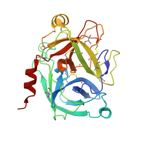
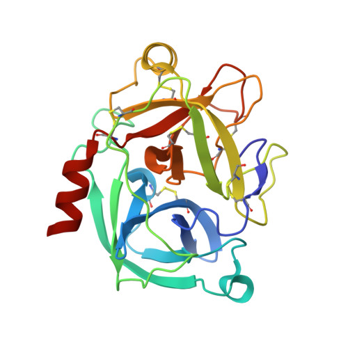
6NVB

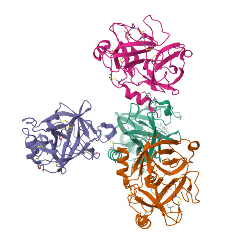
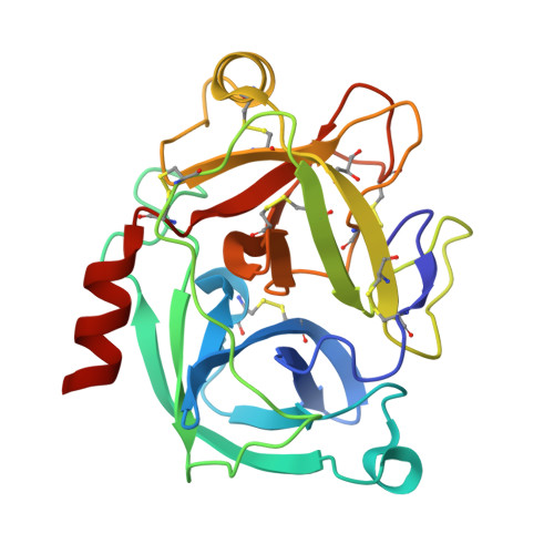
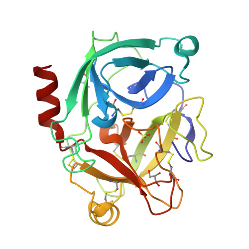
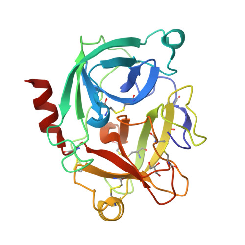
Crystal structure of the inhibitor-free form of the serine protease kallikrein-4


Experimental Data Snapshot

  • Method: X-RAY DIFFRACTION
  • Resolution: 1.64 Å
  • R-Value Free: 0.180 
  • R-Value Work: 0.154 

Starting Model: experimental
View more details

wwPDB Validation   3D Report Full Report


This is version 1.4 of the entry. See complete history


Literature

Crystal structure of the inhibitor-free form of the serine protease kallikrein-4.

Riley, B.T.Hoke, D.E.McGowan, S.Buckle, A.M.

(2019) Acta Crystallogr F Struct Biol Commun 75: 543-546

  • DOI: https://doi.org/10.1107/S2053230X19009610
  • Primary Citation of Related Structures:  
    6NVB

  • PubMed Abstract: 

    Kallikrein 4 (KLK4) is a serine protease that is predominantly expressed in the prostate and is overexpressed in prostate cancer. As such, it has gained attention as an attractive target for prostate cancer therapeutics. Currently, only liganded structures of KLK4 exist in the Protein Data Bank. Until now, inferences about the subtle structural changes in KLK4 upon ligand binding have been made by comparison to other liganded forms, rather than to an apo form. In this study, an inhibitor-free form of KLK4 was crystallized. The crystals obtained belonged to space group P1, contained four molecules in the asymmetric unit and diffracted to 1.64 Å resolution. Interestingly, a nonstandard rotamer of the specificity-determining residue Asp189 was observed in all chains. This model will provide a useful unliganded structure for the future structure-guided design of KLK4 inhibitors.


  • Organizational Affiliation

    Biomedicine Discovery Institute and Department of Biochemistry and Molecular Biology, Monash University, 23 Innovation Walk, Clayton, VIC 3800, Australia.


Macromolecules
Find similar proteins by:  (by identity cutoff)  |  3D Structure
Entity ID: 1
MoleculeChains Sequence LengthOrganismDetailsImage
Kallikrein-4
A, B, C, D
224Homo sapiensMutation(s): 1 
Gene Names: KLK4EMSP1PRSS17PSTS
EC: 3.4.21
UniProt & NIH Common Fund Data Resources
Find proteins for Q9Y5K2 (Homo sapiens)
Explore Q9Y5K2 
Go to UniProtKB:  Q9Y5K2
PHAROS:  Q9Y5K2
GTEx:  ENSG00000167749 
Entity Groups  
Sequence Clusters30% Identity50% Identity70% Identity90% Identity95% Identity100% Identity
UniProt GroupQ9Y5K2
Sequence Annotations
Expand
  • Reference Sequence
Small Molecules
Ligands 2 Unique
IDChains Name / Formula / InChI Key2D Diagram3D Interactions
SO4
Query on SO4

Download Ideal Coordinates CCD File 
H [auth B],
L [auth D]
SULFATE ION
O4 S
QAOWNCQODCNURD-UHFFFAOYSA-L
GOL
Query on GOL

Download Ideal Coordinates CCD File 
E [auth A]
F [auth B]
G [auth B]
I [auth C]
J [auth D]
E [auth A],
F [auth B],
G [auth B],
I [auth C],
J [auth D],
K [auth D]
GLYCEROL
C3 H8 O3
PEDCQBHIVMGVHV-UHFFFAOYSA-N
Experimental Data & Validation

Experimental Data

Unit Cell:
Length ( Å )Angle ( ˚ )
a = 51.602α = 79.01
b = 65.723β = 72.91
c = 74.191γ = 77.62
Software Package:
Software NamePurpose
Blu-Icedata collection
XDSdata reduction
Aimlessdata scaling
PHASERphasing
Cootmodel building
PHENIXrefinement

Structure Validation

View Full Validation Report


Entry History 

Deposition Data

Revision History  (Full details and data files)

  • Version 1.0: 2019-07-17
    Type: Initial release
  • Version 1.1: 2019-08-21
    Changes: Data collection, Database references
  • Version 1.2: 2019-08-28
    Changes: Data collection, Structure summary
  • Version 1.3: 2023-10-11
    Changes: Data collection, Database references, Refinement description
  • Version 1.4: 2024-11-20
    Changes: Structure summary