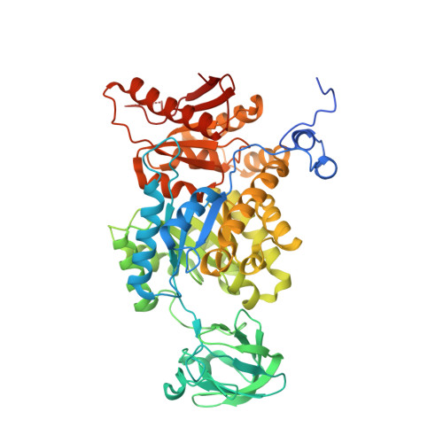
6NUB

Pyruvate Kinase M2 Mutant - S437Y in Complex with L-serine


Experimental Data Snapshot

  • Method: X-RAY DIFFRACTION
  • Resolution: 1.70 Å
  • R-Value Free: 0.174 
  • R-Value Work: 0.154 
  • R-Value Observed: 0.155 

Starting Model: experimental
View more details

wwPDB Validation   3D Report Full Report


This is version 1.3 of the entry. See complete history


Literature

Mechanistic and Structural Insights into Cysteine-Mediated Inhibition of Pyruvate Kinase Muscle Isoform 2.

Srivastava, D.Nandi, S.Dey, M.

(2019) Biochemistry 58: 3669-3682

  • DOI: https://doi.org/10.1021/acs.biochem.9b00349
  • Primary Citation of Related Structures:  
    6NU1, 6NU5, 6NUB

  • PubMed Abstract: 

    Cancer cells regulate key enzymes in the glycolytic pathway to control the glycolytic flux, which is necessary for their growth and proliferation. One of the enzymes is pyruvate kinase muscle isoform 2 (PKM2), which is allosterically regulated by various small molecules. Using detailed biochemical and kinetic studies, we demonstrate that cysteine inhibits wild-type (wt) PKM2 by shifting from an active tetramer to a mixture of a tetramer and a less active dimer/monomer equilibrium and that the inhibition is dependent on cysteine concentration. The cysteine-mediated PKM2 inhibition is reversed by fructose 1,6-bisphosphate, an allosteric activator of PKM2. Furthermore, kinetic studies using two dimeric PKM2 variants, S437Y PKM2 and G415R PKM2, show that the reversal is caused by the tetramerization of wtPKM2. The crystal structure of the wtPKM2-Cys complex was determined at 2.25 Å, which showed that cysteine is held to the amino acid binding site via its main chain groups, similar to that observed for phenylalanine, alanine, serine, and tryptophan. Notably, ligand binding studies using fluorescence and isothermal titration calorimetry show that the presence of phosphoenolpyruvate alters the binding affinities of amino acids for wtPKM2 and vice versa, thereby unravelling the existence of a functionally bidirectional coupling between the amino acid binding site and the active site of wtPKM2.


  • Organizational Affiliation

    Department of Chemistry , The University of Iowa , Iowa City , Iowa 52242 , United States.


Macromolecules
Find similar proteins by:  (by identity cutoff)  |  3D Structure
Entity ID: 1
MoleculeChains Sequence LengthOrganismDetailsImage
Pyruvate kinase PKM
A, B
550Homo sapiensMutation(s): 1 
Gene Names: PKMOIP3PK2PK3PKM2
EC: 2.7.1.40 (PDB Primary Data), 2.7.11.1 (UniProt), 2.7.10.2 (UniProt)
UniProt & NIH Common Fund Data Resources
Find proteins for P14618 (Homo sapiens)
Explore P14618 
Go to UniProtKB:  P14618
PHAROS:  P14618
GTEx:  ENSG00000067225 
Entity Groups  
Sequence Clusters30% Identity50% Identity70% Identity90% Identity95% Identity100% Identity
UniProt GroupP14618
Sequence Annotations
Expand
  • Reference Sequence
Small Molecules
Ligands 9 Unique
IDChains Name / Formula / InChI Key2D Diagram3D Interactions
B3P
Query on B3P

Download Ideal Coordinates CCD File 
J [auth A],
T [auth B]
2-[3-(2-HYDROXY-1,1-DIHYDROXYMETHYL-ETHYLAMINO)-PROPYLAMINO]-2-HYDROXYMETHYL-PROPANE-1,3-DIOL
C11 H26 N2 O6
HHKZCCWKTZRCCL-UHFFFAOYSA-N
PG4
Query on PG4

Download Ideal Coordinates CCD File 
R [auth B]TETRAETHYLENE GLYCOL
C8 H18 O5
UWHCKJMYHZGTIT-UHFFFAOYSA-N
PEG
Query on PEG

Download Ideal Coordinates CCD File 
I [auth A]DI(HYDROXYETHYL)ETHER
C4 H10 O3
MTHSVFCYNBDYFN-UHFFFAOYSA-N
SER
Query on SER

Download Ideal Coordinates CCD File 
C [auth A],
K [auth B]
SERINE
C3 H7 N O3
MTCFGRXMJLQNBG-REOHCLBHSA-N
OXL
Query on OXL

Download Ideal Coordinates CCD File 
E [auth A],
M [auth B]
OXALATE ION
C2 O4
MUBZPKHOEPUJKR-UHFFFAOYSA-L
EDO
Query on EDO

Download Ideal Coordinates CCD File 
S [auth B]1,2-ETHANEDIOL
C2 H6 O2
LYCAIKOWRPUZTN-UHFFFAOYSA-N
SCN
Query on SCN

Download Ideal Coordinates CCD File 
G [auth A],
H [auth A],
O [auth B],
P [auth B],
Q [auth B]
THIOCYANATE ION
C N S
ZMZDMBWJUHKJPS-UHFFFAOYSA-M
K
Query on K

Download Ideal Coordinates CCD File 
F [auth A],
N [auth B]
POTASSIUM ION
K
NPYPAHLBTDXSSS-UHFFFAOYSA-N
MG
Query on MG

Download Ideal Coordinates CCD File 
D [auth A],
L [auth B]
MAGNESIUM ION
Mg
JLVVSXFLKOJNIY-UHFFFAOYSA-N
Experimental Data & Validation

Experimental Data

  • Method: X-RAY DIFFRACTION
  • Resolution: 1.70 Å
  • R-Value Free: 0.174 
  • R-Value Work: 0.154 
  • R-Value Observed: 0.155 
  • Space Group: C 1 2 1
Unit Cell:
Length ( Å )Angle ( ˚ )
a = 110.11α = 90
b = 94.23β = 95.63
c = 109.13γ = 90
Software Package:
Software NamePurpose
PHENIXrefinement
XDSdata reduction
SCALAdata scaling
PHASERphasing
Cootmodel building

Structure Validation

View Full Validation Report


Entry History & Funding Information

Deposition Data


Funding OrganizationLocationGrant Number
National Science Foundation (NSF, United States)United StatesCLP 1506181

Revision History  (Full details and data files)

  • Version 1.0: 2019-08-21
    Type: Initial release
  • Version 1.1: 2019-09-18
    Changes: Data collection, Database references
  • Version 1.2: 2019-11-27
    Changes: Author supporting evidence
  • Version 1.3: 2023-10-11
    Changes: Advisory, Data collection, Database references, Derived calculations, Refinement description