


Crystal structure of Mycobacterium tuberculosis dethiobiotin synthetase in complex with fragment analogue 5
Thompson, A.P., Polyak, S.W., Wegener, K.L., Bruning, J.B.To be published.
Experimental Data Snapshot
Starting Model: experimental
View more details
Entity ID: 1 | |||||
|---|---|---|---|---|---|
| Molecule | Chains | Sequence Length | Organism | Details | Image |
| ATP-dependent dethiobiotin synthetase BioD | 233 | Mycobacterium tuberculosis H37Rv | Mutation(s): 0 Gene Names: bioD, Rv1570, MTCY336.33c EC: 6.3.3.3 | 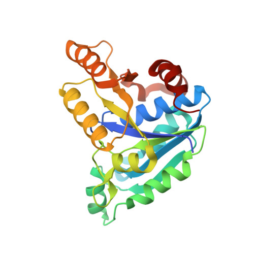 | |
UniProt | |||||
Entity Groups | |||||
| Sequence Clusters | 30% Identity50% Identity70% Identity90% Identity95% Identity100% Identity | ||||
| UniProt Group | P9WPQ5 | ||||
Sequence AnnotationsExpand | |||||
Reference Sequence | |||||
| Ligands 3 Unique | |||||
|---|---|---|---|---|---|
| ID | Chains | Name / Formula / InChI Key | 2D Diagram | 3D Interactions | |
| L1V (Subject of Investigation/LOI) Download:Ideal Coordinates CCD File | E [auth A], H [auth B], K [auth C], N [auth D] | 4-[(1R,2S)-2-(carboxymethyl)cyclopentane-1-carbonyl]benzoic acid C15 H16 O5 AIBKYDBZFDNIIN-NWDGAFQWSA-N |  | ||
| L1Y (Subject of Investigation/LOI) Download:Ideal Coordinates CCD File | F [auth A], I [auth B], L [auth C], O [auth D] | 4-[(1S,2R)-2-(carboxymethyl)cyclopentane-1-carbonyl]benzoic acid C15 H16 O5 AIBKYDBZFDNIIN-NEPJUHHUSA-N |  | ||
| SO4 (Subject of Investigation/LOI) Download:Ideal Coordinates CCD File | G [auth A], J [auth B], M [auth C], P [auth D] | SULFATE ION O4 S QAOWNCQODCNURD-UHFFFAOYSA-L |  | ||
| Length ( Å ) | Angle ( ˚ ) |
|---|---|
| a = 54.797 | α = 90 |
| b = 105.593 | β = 90 |
| c = 152.523 | γ = 90 |
| Software Name | Purpose |
|---|---|
| XDS | data reduction |
| Aimless | data scaling |
| PHASER | phasing |
| PHENIX | refinement |
| PDB_EXTRACT | data extraction |
| Funding Organization | Location | Grant Number |
|---|---|---|
| National Health and Medical Research Council (NHMRC, Australia) | Australia | APP1068885 |


