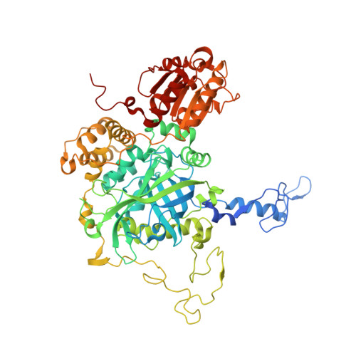
X-ray driven reduction of Cpd I of Catalase-3 from N. crassa reveals differential sensitivity of active sites and formation of ferrous state.
Zarate-Romero, A., Stojanoff, V., Cohen, A.E., Hansberg, W., Rudino-Pinera, E.(2019) Arch Biochem Biophys 666: 107-115
- PubMed: 30940570
- DOI: https://doi.org/10.1016/j.abb.2019.03.020
- Primary Citation of Related Structures:
4AJ9, 6NSW, 6NSY, 6NSZ, 6NT0, 6NT1 - PubMed Abstract:
Catalases are biotechnologically relevant enzymes because of their applications in food technology, bioremediation, and biomedicine. The dismutation of hydrogen peroxide occurs in two steps; in the first one, the enzyme forms an oxidized compound I (Cpd I) and in the second one, the enzyme is reduced to the ferric state. In this research work, we analyzed the reduction of Cpd I by X-ray radiation damage during diffraction experiments in crystals of CAT-3, a Large-Size Subunit Catalase (LSC) from Neurospora crassa. A Multi-Crystal Data collection Strategy was applied in order to obtain the Cpd I structure at a resolution of 2.2 Å; this intermediate was highly sensitive to X-ray and was easily reduced at very low deposited radiation dose, causing breakage of the Fe=O bond. The comparison of the structures showed reduced intermediates and also evidenced the differential sensitivity per monomer. The resting ferric state was reduced to the ferrous state, an intermediate without a previous report in LSC. The chemically obtained Cpd I and the X-ray reduced intermediates were identified by UV-visible microspectrometry coupled to data collection. The differential sensitivity and the formation of a ferrous state are discussed, emphasizing the importance of the correct interpretation in the oxidation state of the iron heme.
Organizational Affiliation:
Cátedras CONACyT, Departamento de Bionanotecnología, Centro de Nanociencias y Nanotecnología, Universidad Nacional Autónoma de México, Ensenada, B. C., Mexico.


