6NNH | pdb_00006nnh

Structure of Closed state of Dihydrofolate reductase from Mycobacterium tuberculosis in complex with NADPH and cycloguanil


Experimental Data Snapshot

  • Method: X-RAY DIFFRACTION
  • Resolution: 1.52 Å
  • R-Value Free: 
    0.199 (Depositor), 0.201 (DCC) 
  • R-Value Work: 
    0.170 (Depositor) 
  • R-Value Observed: 
    0.171 (Depositor) 

Starting Model: experimental
View more details

wwPDB Validation 3D Report Full Report

Validation slider image for 6NNH

Ligand Structure Quality Assessment 


This is version 1.2 of the entry. See complete history

Literature

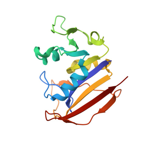
Crystal structures of the closed form of Mycobacterium tuberculosis dihydrofolate reductase in complex with dihydrofolate and antifolates.

Ribeiro, J.A.Chavez-Pacheco, S.M.de Oliveira, G.S.Silva, C.D.S.Giudice, J.H.P.Libreros-Zuniga, G.A.Dias, M.V.B.

(2019) Acta Crystallogr D Struct Biol 75: 682-693

  • DOI: https://doi.org/10.1107/S205979831900901X
  • Primary Citation Related Structures: 
    6NNC, 6NND, 6NNE, 6NNH, 6NNI

  • PubMed Abstract: 

    Tuberculosis is a disease caused by Mycobacterium tuberculosis and is the leading cause of death from a single infectious pathogen, with a high prevalence in developing countries in Africa and Asia. There still is a need for the development or repurposing of novel therapies to combat this disease owing to the long-term nature of current therapies and because of the number of reported resistant strains. Here, structures of dihydrofolate reductase from M. tuberculosis (MtDHFR), which is a key target of the folate pathway, are reported in complex with four antifolates, pyrimethamine, cycloguanil, diaverdine and pemetrexed, and its substrate dihydrofolate in order to understand their binding modes. The structures of all of these complexes were obtained in the closed-conformation state of the enzyme and a fine structural analysis indicated motion in key regions of the substrate-binding site and different binding modes of the ligands. In addition, the affinities, through K d measurement, of diaverdine and methotrexate have been determined; MtDHFR has a lower affinity (highest K d ) for diaverdine than pyrimethamine and trimethoprim, and a very high affinity for methotrexate, as expected. The structural comparisons and analysis described in this work provide new information about the plasticity of MtDHFR and the binding effects of different antifolates.


  • Organizational Affiliation
    • Department of Microbiology, Institute of Biomedical Science, University of São Paulo, Avenida Professor Lineu Prestes 1374, São Paulo 05508-000, Brazil.

Macromolecule Content 

  • Total Structure Weight: 38.01 kDa 
  • Atom Count: 3,011 
  • Modeled Residue Count: 318 
  • Deposited Residue Count: 318 
  • Unique protein chains: 1

Macromolecules

Find similar proteins by:|  3D Structure
Entity ID: 1
MoleculeChains  Sequence LengthOrganismDetailsImage
Dihydrofolate reductase
A, B
159Mycobacterium tuberculosis H37RvMutation(s): 0 
Gene Names: folAdfrARv2763cMTV002.28c
EC: 1.5.1.3
UniProt
Find proteins for P9WNX1 (Mycobacterium tuberculosis (strain ATCC 25618 / H37Rv))
Explore P9WNX1 
Go to UniProtKB:  P9WNX1
Entity Groups
Sequence Clusters30% Identity50% Identity70% Identity90% Identity95% Identity100% Identity
UniProt GroupP9WNX1
Sequence Annotations
Expand
Reference Sequence

Small Molecules

Ligands 5 Unique
IDChains Name / Formula / InChI Key2D Diagram3D Interactions
NDP

Query on NDP


Download:Ideal Coordinates CCD File
D [auth A],
J [auth B]
NADPH DIHYDRO-NICOTINAMIDE-ADENINE-DINUCLEOTIDE PHOSPHATE
C21 H30 N7 O17 P3
ACFIXJIJDZMPPO-NNYOXOHSSA-N
1CY
(Subject of Investigation/LOI)

Query on 1CY


Download:Ideal Coordinates CCD File
C [auth A],
I [auth B]
1-(4-chlorophenyl)-6,6-dimethyl-1,6-dihydro-1,3,5-triazine-2,4-diamine
C11 H14 Cl N5
QMNFFXRFOJIOKZ-UHFFFAOYSA-N
SO4

Query on SO4


Download:Ideal Coordinates CCD File
E [auth A],
F [auth A],
G [auth A],
K [auth B],
L [auth B]
SULFATE ION
O4 S
QAOWNCQODCNURD-UHFFFAOYSA-L
GOL

Query on GOL


Download:Ideal Coordinates CCD File
N [auth B]GLYCEROL
C3 H8 O3
PEDCQBHIVMGVHV-UHFFFAOYSA-N
CO

Query on CO


Download:Ideal Coordinates CCD File
H [auth A],
M [auth B]
COBALT (II) ION
Co
XLJKHNWPARRRJB-UHFFFAOYSA-N

Experimental Data & Validation

Experimental Data

  • Method: X-RAY DIFFRACTION
  • Resolution: 1.52 Å
  • R-Value Free:  0.199 (Depositor), 0.201 (DCC) 
  • R-Value Work:  0.170 (Depositor) 
  • R-Value Observed: 0.171 (Depositor) 
Space Group: P 21 21 21
Unit Cell:
Length ( Å )Angle ( ˚ )
a = 60.319α = 90
b = 71.245β = 90
c = 72.219γ = 90
Software Package:
Software NamePurpose
PHENIXrefinement
Aimlessdata scaling
PHASERphasing
PDB_EXTRACTdata extraction
XDSdata reduction

Structure Validation

View Full Validation Report


Ligand Structure Quality Assessment 


Entry History 

Deposition Data

Revision History  (Full details and data files)

  • Version 1.0: 2019-07-17
    Type: Initial release
  • Version 1.1: 2019-11-13
    Changes: Database references
  • Version 1.2: 2023-10-11
    Changes: Data collection, Database references, Derived calculations, Refinement description