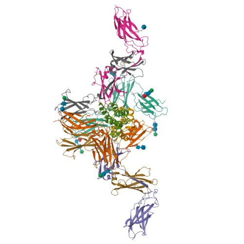
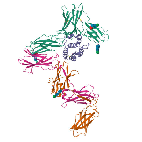
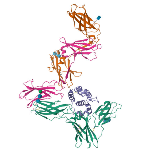
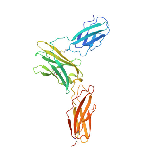
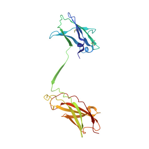
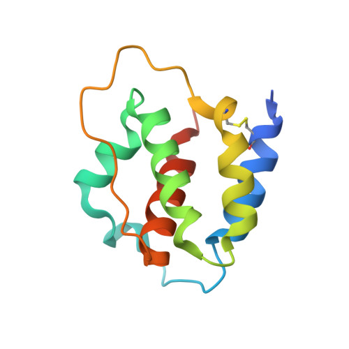
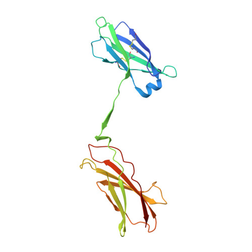
6NMY

A Cytokine-receptor complex


Experimental Data Snapshot

  • Method: X-RAY DIFFRACTION
  • Resolution: 3.30 Å
  • R-Value Free: 0.278 
  • R-Value Work: 0.236 
  • R-Value Observed: 0.238 

Starting Models: experimental
View more details

wwPDB Validation   3D Report Full Report


Ligand Structure Quality Assessment 


This is version 2.2 of the entry. See complete history


Literature

Distinct Assemblies of Heterodimeric Cytokine Receptors Govern Stemness Programs in Leukemia.

Kan, W.L.Dhagat, U.Kaufmann, K.B.Hercus, T.R.Nero, T.L.Zeng, A.G.X.Toubia, J.Barry, E.F.Broughton, S.E.Gomez, G.A.Benard, B.A.Dottore, M.Cheung Tung Shing, K.S.Boutzen, H.Samaraweera, S.E.Simpson, K.J.Jin, L.Goodall, G.J.Begley, C.G.Thomas, D.Ekert, P.G.Tvorogov, D.D'Andrea, R.J.Dick, J.E.Parker, M.W.Lopez, A.F.

(2023) Cancer Discov 13: 1922-1947

  • DOI: https://doi.org/10.1158/2159-8290.CD-22-1396
  • Primary Citation of Related Structures:  
    6NMY

  • PubMed Abstract: 

    Leukemia stem cells (LSC) possess distinct self-renewal and arrested differentiation properties that are responsible for disease emergence, therapy failure, and recurrence in acute myeloid leukemia (AML). Despite AML displaying extensive biological and clinical heterogeneity, LSC with high interleukin-3 receptor (IL3R) levels are a constant yet puzzling feature, as this receptor lacks tyrosine kinase activity. Here, we show that the heterodimeric IL3Rα/βc receptor assembles into hexamers and dodecamers through a unique interface in the 3D structure, where high IL3Rα/βc ratios bias hexamer formation. Importantly, receptor stoichiometry is clinically relevant as it varies across the individual cells in the AML hierarchy, in which high IL3Rα/βc ratios in LSCs drive hexamer-mediated stemness programs and poor patient survival, while low ratios mediate differentiation. Our study establishes a new paradigm in which alternative cytokine receptor stoichiometries differentially regulate cell fate, a signaling mechanism that may be generalizable to other transformed cellular hierarchies and of potential therapeutic significance. Stemness is a hallmark of many cancers and is largely responsible for disease emergence, progression, and relapse. Our finding that clinically significant stemness programs in AML are directly regulated by different stoichiometries of cytokine receptors represents a hitherto unexplained mechanism underlying cell-fate decisions in cancer stem cell hierarchies. This article is highlighted in the In This Issue feature, p. 1749.


  • Organizational Affiliation

    Cytokine Receptor Laboratory, Centre for Cancer Biology, SA Pathology and the University of South Australia, Adelaide, South Australia, Australia.


Macromolecules
Find similar proteins by:  (by identity cutoff)  |  3D Structure
Entity ID: 1
MoleculeChains Sequence LengthOrganismDetailsImage
Interleukin-3 receptor subunit alphaA [auth F],
B [auth M]
288Homo sapiensMutation(s): 1 
Gene Names: IL3RAIL3R
UniProt & NIH Common Fund Data Resources
Find proteins for P26951 (Homo sapiens)
Explore P26951 
Go to UniProtKB:  P26951
PHAROS:  P26951
GTEx:  ENSG00000185291 
Entity Groups  
Sequence Clusters30% Identity50% Identity70% Identity90% Identity95% Identity100% Identity
UniProt GroupP26951
Glycosylation
Glycosylation Sites: 3Go to GlyGen: P26951-1
Sequence Annotations
Expand
  • Reference Sequence
Find similar proteins by:  (by identity cutoff)  |  3D Structure
Entity ID: 2
MoleculeChains Sequence LengthOrganismDetailsImage
Cytokine receptor common subunit betaC [auth A],
D [auth C]
216Homo sapiensMutation(s): 0 
Gene Names: CSF2RBIL3RBIL5RB
UniProt & NIH Common Fund Data Resources
Find proteins for P32927 (Homo sapiens)
Explore P32927 
Go to UniProtKB:  P32927
PHAROS:  P32927
GTEx:  ENSG00000100368 
Entity Groups  
Sequence Clusters30% Identity50% Identity70% Identity90% Identity95% Identity100% Identity
UniProt GroupP32927
Glycosylation
Glycosylation Sites: 1Go to GlyGen: P32927-1
Sequence Annotations
Expand
  • Reference Sequence
Find similar proteins by:  (by identity cutoff)  |  3D Structure
Entity ID: 3
MoleculeChains Sequence LengthOrganismDetailsImage
Interleukin-3E [auth I],
F [auth J]
118Homo sapiensMutation(s): 1 
Gene Names: IL3
UniProt & NIH Common Fund Data Resources
Find proteins for P08700 (Homo sapiens)
Explore P08700 
Go to UniProtKB:  P08700
PHAROS:  P08700
GTEx:  ENSG00000164399 
Entity Groups  
Sequence Clusters30% Identity50% Identity70% Identity90% Identity95% Identity100% Identity
UniProt GroupP08700
Sequence Annotations
Expand
  • Reference Sequence
Find similar proteins by:  (by identity cutoff)  |  3D Structure
Entity ID: 4
MoleculeChains Sequence LengthOrganismDetailsImage
Cytokine receptor common subunit betaG [auth B],
H [auth D]
198Homo sapiensMutation(s): 1 
Gene Names: CSF2RBIL3RBIL5RB
UniProt & NIH Common Fund Data Resources
Find proteins for P32927 (Homo sapiens)
Explore P32927 
Go to UniProtKB:  P32927
PHAROS:  P32927
GTEx:  ENSG00000100368 
Entity Groups  
Sequence Clusters30% Identity50% Identity70% Identity90% Identity95% Identity100% Identity
UniProt GroupP32927
Sequence Annotations
Expand
  • Reference Sequence
Oligosaccharides

Help

Entity ID: 5
MoleculeChains Length2D Diagram Glycosylation3D Interactions
beta-D-mannopyranose-(1-4)-2-acetamido-2-deoxy-beta-D-glucopyranose-(1-4)-[alpha-L-fucopyranose-(1-6)]2-acetamido-2-deoxy-beta-D-glucopyranoseI [auth E],
J [auth G]
4N-Glycosylation
Glycosylation Resources
GlyTouCan:  G32152BH
GlyCosmos:  G32152BH
GlyGen:  G32152BH
Entity ID: 6
MoleculeChains Length2D Diagram Glycosylation3D Interactions
beta-D-mannopyranose-(1-4)-2-acetamido-2-deoxy-beta-D-glucopyranose-(1-4)-2-acetamido-2-deoxy-beta-D-glucopyranoseK [auth H],
L [auth K],
N
3N-Glycosylation
Glycosylation Resources
GlyTouCan:  G15407YE
GlyCosmos:  G15407YE
GlyGen:  G15407YE
Entity ID: 7
MoleculeChains Length2D Diagram Glycosylation3D Interactions
2-acetamido-2-deoxy-beta-D-glucopyranose-(1-4)-2-acetamido-2-deoxy-beta-D-glucopyranoseM [auth L],
O
2N-Glycosylation
Glycosylation Resources
GlyTouCan:  G42666HT
GlyCosmos:  G42666HT
GlyGen:  G42666HT
Small Molecules
Ligands 1 Unique
IDChains Name / Formula / InChI Key2D Diagram3D Interactions
NAG
Query on NAG

Download Ideal Coordinates CCD File 
P [auth C]2-acetamido-2-deoxy-beta-D-glucopyranose
C8 H15 N O6
OVRNDRQMDRJTHS-FMDGEEDCSA-N
Experimental Data & Validation

Experimental Data

  • Method: X-RAY DIFFRACTION
  • Resolution: 3.30 Å
  • R-Value Free: 0.278 
  • R-Value Work: 0.236 
  • R-Value Observed: 0.238 
  • Space Group: P 21 21 21
Unit Cell:
Length ( Å )Angle ( ˚ )
a = 111.647α = 90
b = 157.251β = 90
c = 168.306γ = 90
Software Package:
Software NamePurpose
PHENIXrefinement
Aimlessdata scaling
PHASERphasing
PDB_EXTRACTdata extraction
XDSdata reduction

Structure Validation

View Full Validation Report


Ligand Structure Quality Assessment 


Entry History & Funding Information

Deposition Data


Funding OrganizationLocationGrant Number
National Health and Medical Research Council (NHMRC, Australia)Australia--

Revision History  (Full details and data files)

  • Version 1.0: 2020-01-22
    Type: Initial release
  • Version 2.0: 2020-07-29
    Type: Remediation
    Reason: Carbohydrate remediation
    Changes: Advisory, Atomic model, Data collection, Derived calculations, Structure summary
  • Version 2.1: 2023-10-11
    Changes: Data collection, Database references, Derived calculations, Refinement description, Structure summary
  • Version 2.2: 2024-10-16
    Changes: Database references, Structure summary