6NMA | pdb_00006nma

CryoEM structure of the LbCas12a-crRNA-AcrVA4 complex


Experimental Data Snapshot

  • Method: ELECTRON MICROSCOPY
  • Resolution: 3.38 Å
  • Aggregation State: PARTICLE 
  • Reconstruction Method: SINGLE PARTICLE 

wwPDB Validation 3D Report Full Report

Validation slider image for 6NMA

This is version 1.3 of the entry. See complete history

Literature

Structural Basis for the Inhibition of CRISPR-Cas12a by Anti-CRISPR Proteins.

Zhang, H.Li, Z.Daczkowski, C.M.Gabel, C.Mesecar, A.D.Chang, L.

(2019) Cell Host Microbe 25: 815

  • DOI: https://doi.org/10.1016/j.chom.2019.05.004
  • Primary Citation Related Structures: 
    6NM9, 6NMA, 6NMC, 6NMD, 6NME, 6OMV

  • PubMed Abstract: 

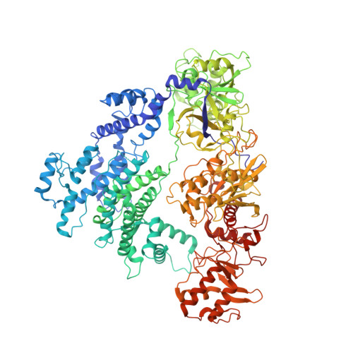
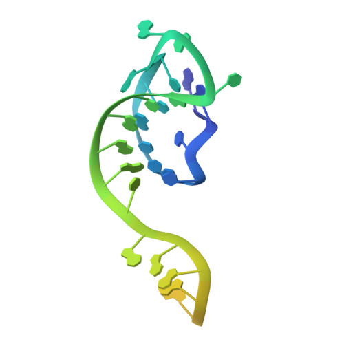
    CRISPR-Cas12a (Cpf1), a type V CRISPR-associated nuclease, provides bacterial immunity against bacteriophages and plasmids but also serves as a tool for genome editing. Foreign nucleic acids are integrated into the CRISPR locus, prompting transcription of CRISPR RNAs (crRNAs) that guide Cas12a cleavage of foreign complementary DNA. However, mobile genetic elements counteract Cas12a with inhibitors, notably type V-A anti-CRISPRs (AcrVAs). We present cryoelectron microscopy structures of Cas12a-crRNA bound to AcrVA1 and AcrVA4 at 3.5 and 3.3 Å resolutions, respectively. AcrVA1 is sandwiched between the recognition (REC) and nuclease (NUC) lobes of Cas12a and inserts into the binding pocket for the protospacer-adjacent motif (PAM), a short DNA sequence guiding Cas12a targeting. AcrVA1 cleaves crRNA in a Cas12a-dependent manner, inactivating Cas12a-crRNA complexes. The AcrVA4 dimer is anchored around the crRNA pseudoknot of Cas12a-crRNA, preventing required conformational changes for crRNA-DNA heteroduplex formation. These results uncover molecular mechanisms for CRISPR-Cas12a inhibition, providing insights into bacteria-phage dynamics.


  • Organizational Affiliation
    • Department of Biological Sciences, Purdue University, West Lafayette, IN 47907, USA.

Macromolecule Content 

  • Total Structure Weight: 211.42 kDa 
  • Atom Count: 12,404 
  • Modeled Residue Count: 1,460 
  • Deposited Residue Count: 1,735 
  • Unique protein chains: 2
  • Unique nucleic acid chains: 1

Macromolecules


Find similar proteins by:|  3D Structure
Entity ID: 1
MoleculeChains  Sequence LengthOrganismDetailsImage
AcrVA1A,
D [auth C]
234Moraxella bovoculiMutation(s): 0 
Gene Names: AAX07_09545
Find similar proteins by:|  3D Structure
Entity ID: 2
MoleculeChains  Sequence LengthOrganismDetailsImage
Cpf11,227Lachnospiraceae bacterium ND2006Mutation(s): 0 
UniProt
Find proteins for A0ACD6BAZ6 (Lachnospiraceae bacterium ND2006)
Explore A0ACD6BAZ6 
Go to UniProtKB:  A0ACD6BAZ6
Entity Groups
Sequence Clusters30% Identity50% Identity70% Identity90% Identity95% Identity100% Identity
UniProt GroupA0ACD6BAZ6
Sequence Annotations
Expand
Reference Sequence
Find similar nucleic acids by:  (by identity cutoff) 
Entity ID: 3
MoleculeChains LengthOrganismImage
RNAC [auth G]40Lachnospiraceae bacterium ND2006
Sequence Annotations
Expand
Reference Sequence

Experimental Data & Validation

Experimental Data

  • Method: ELECTRON MICROSCOPY
  • Resolution: 3.38 Å
  • Aggregation State: PARTICLE 
  • Reconstruction Method: SINGLE PARTICLE 
EM Software:
TaskSoftware PackageVersion
RECONSTRUCTIONcisTEM1.0.0

Structure Validation

View Full Validation Report


Entry History 

Deposition Data

Revision History  (Full details and data files)

  • Version 1.0: 2019-06-12
    Type: Initial release
  • Version 1.1: 2019-06-26
    Changes: Data collection, Database references
  • Version 1.2: 2019-12-18
    Changes: Other
  • Version 1.3: 2024-03-20
    Changes: Data collection, Database references