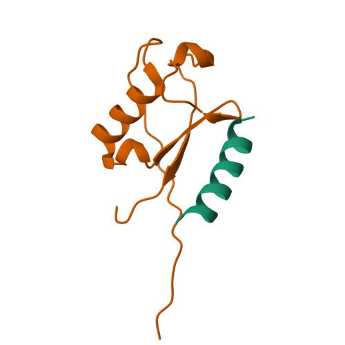
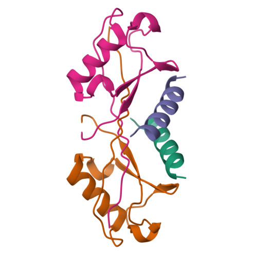
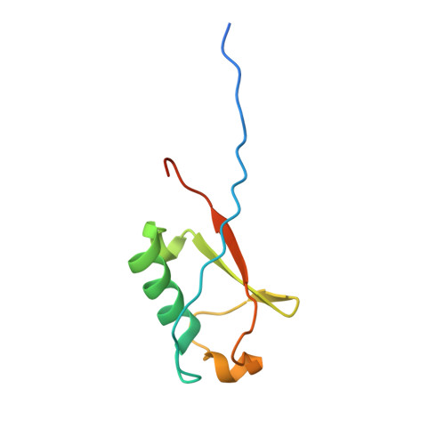
6NJG

Ubiquitin Variant in Complex with Ubiquitin Interacting Motif


Experimental Data Snapshot

  • Method: X-RAY DIFFRACTION
  • Resolution: 2.35 Å
  • R-Value Free: 0.244 
  • R-Value Work: 0.206 
  • R-Value Observed: 0.209 

Starting Models: experimental
View more details

wwPDB Validation   3D Report Full Report


This is version 1.3 of the entry. See complete history


Literature

Dimerization of a ubiquitin variant leads to high affinity interactions with a ubiquitin interacting motif.

Manczyk, N.Veggiani, G.Gish, G.D.Yates, B.P.Ernst, A.Sidhu, S.S.Sicheri, F.

(2019) Protein Sci 28: 848-856

  • DOI: https://doi.org/10.1002/pro.3593
  • Primary Citation of Related Structures:  
    6NJG

  • PubMed Abstract: 

    We previously described structural and functional characterization of the first ubiquitin variant (UbV), UbV.v27.1, engineered by phage display to bind with high affinity to a specific ubiquitin interacting motif (UIM). We identified two substitutions relative to ubiquitin (Gly10Val/His68Tyr) that were critical for enhancing binding affinity but could only rationalize the mechanism of action of the Tyr68 substitution. Here, we extend our characterization and uncover the mechanism by which the Val10 substitution enhances binding affinity. We show that Val10 in UbV.v27.1 drives UbV dimerization through an intermolecular β-strand exchange. Dimerization serves to increase the contact surface between the UIM and UbV and also affords direct contacts between two UIMs through an overall 2:2 binding stoichiometry. Our identification of the role of Val10 in UbV dimerization suggests a general means for the development of dimeric UbVs with improved affinity and specificity relative to their monomeric UbV counterparts. Statement: Previously, we used phage display to engineer a UbV that bound tightly and specifically to a UIM. Here, we discovered that tight binding is partly due to the dimerization of the UbV, which increases the contact surface between the UbV and UIM. We show that UbV dimerization is dependent on the Gly10Val substitution, and posit that dimerization may provide a general means for engineering UbVs with improved binding properties.


  • Organizational Affiliation

    Lunenfeld-Tanenbaum Research Institute, Mount Sinai Hospital, Toronto, Ontario M5G 1X5, Canada.


Macromolecules

Find similar proteins by:  Sequence   |   3D Structure  

Entity ID: 1
MoleculeChains Sequence LengthOrganismDetailsImage
Vacuolar protein sorting-associated protein 27A [auth B]24Saccharomyces cerevisiae S288CMutation(s): 0 
UniProt
Find proteins for P40343 (Saccharomyces cerevisiae (strain ATCC 204508 / S288c))
Explore P40343 
Go to UniProtKB:  P40343
Entity Groups  
Sequence Clusters30% Identity50% Identity70% Identity90% Identity95% Identity100% Identity
UniProt GroupP40343
Sequence Annotations
Expand
  • Reference Sequence
Find similar proteins by:  (by identity cutoff)  |  3D Structure
Entity ID: 2
MoleculeChains Sequence LengthOrganismDetailsImage
Polyubiquitin-BB [auth C]89Homo sapiensMutation(s): 18 
Gene Names: UBB
UniProt & NIH Common Fund Data Resources
Find proteins for J3QS39 (Homo sapiens)
Explore J3QS39 
Go to UniProtKB:  J3QS39
GTEx:  ENSG00000170315 
Entity Groups  
Sequence Clusters30% Identity50% Identity70% Identity90% Identity95% Identity100% Identity
UniProt GroupJ3QS39
Sequence Annotations
Expand
  • Reference Sequence
Experimental Data & Validation

Experimental Data

  • Method: X-RAY DIFFRACTION
  • Resolution: 2.35 Å
  • R-Value Free: 0.244 
  • R-Value Work: 0.206 
  • R-Value Observed: 0.209 
  • Space Group: P 43 21 2
Unit Cell:
Length ( Å )Angle ( ˚ )
a = 44.613α = 90
b = 44.613β = 90
c = 104.343γ = 90
Software Package:
Software NamePurpose
PHENIXrefinement
REFMACrefinement
HKL-2000data reduction
HKL-2000data scaling
PHASERphasing

Structure Validation

View Full Validation Report


Entry History & Funding Information

Deposition Data


Funding OrganizationLocationGrant Number
Canadian Institutes of Health Research (CIHR)CanadaFDN-143277

Revision History  (Full details and data files)

  • Version 1.0: 2019-03-06
    Type: Initial release
  • Version 1.1: 2019-04-24
    Changes: Data collection, Database references
  • Version 1.2: 2020-01-08
    Changes: Author supporting evidence
  • Version 1.3: 2023-10-11
    Changes: Data collection, Database references, Refinement description