6NHK | pdb_00006nhk

Mortalin nucleotide binding domain in the ADP-bound state

  • Classification: CHAPERONE
  • Organism(s): Homo sapiens
  • Expression System: Escherichia coli
  • Mutation(s): No 

  • Deposited: 2018-12-23 Released: 2019-04-17 
  • Deposition Author(s): Page, R.C., Moseng, M.A., Nix, J.C.
  • Funding Organization(s): National Institutes of Health/National Institute Of Allergy and Infectious Diseases (NIH/NIAID), National Science Foundation (NSF, United States)

Experimental Data Snapshot

  • Method: X-RAY DIFFRACTION
  • Resolution: 2.78 Å
  • R-Value Free: 
    0.296 (Depositor), 0.298 (DCC) 
  • R-Value Work: 
    0.256 (Depositor), 0.260 (DCC) 
  • R-Value Observed: 
    0.259 (Depositor) 

Starting Model: experimental
View more details

wwPDB Validation 3D Report Full Report

Validation slider image for 6NHK

Ligand Structure Quality Assessment 


This is version 1.3 of the entry. See complete history

Literature

Biophysical Consequences of EVEN-PLUS Syndrome Mutations for the Function of Mortalin.

Moseng, M.A.Nix, J.C.Page, R.C.

(2019) J Phys Chem B 123: 3383-3396

  • DOI: https://doi.org/10.1021/acs.jpcb.9b00071
  • Primary Citation Related Structures: 
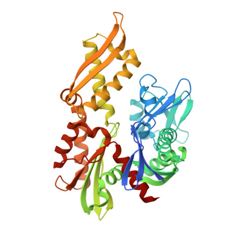
    6NHK

  • PubMed Abstract: 

    HSPA9, the gene coding for the mitochondrial chaperone mortalin, is involved in various cellular roles such as mitochondrial protein import, folding, degradation, Fe-S cluster biogenesis, mitochondrial homeostasis, and regulation of the antiapoptotic protein p53. Mutations in the HSPA9 gene, particularly within the region coding for the nucleotide-binding domain (NBD), cause the autosomal disorder known as EVEN-PLUS syndrome. The resulting mutants R126W and Y128C are located on the surface of the mortalin-NBD near the binding interface with the interdomain linker (IDL). We used differential scanning fluorimetry (DSF), biolayer interferometry, X-ray crystallography, ATP hydrolysis assays, and Rosetta docking simulations to study the structural and functional consequences of the EVEN-PLUS syndrome-associated R126W and Y128C mutations within the mortalin-NBD. These results indicate that the surface mutations R126W and Y128C have far-reaching effects that disrupt ATP hydrolysis, interdomain linker binding, and thermostability and increase the propensity for aggregation. The structural differences observed provide insight into how the conformations of mortalin differ from other heat shock protein 70 (Hsp70) homologues. Combined, our biophysical and structural studies contribute to the understanding of the molecular basis for how disease-associated mortalin mutations affect mortalin functionality and the pathogenesis of EVEN-PLUS syndrome.


  • Organizational Affiliation
    • Department of Chemistry and Biochemistry , Miami University , Oxford , Ohio 45056 , United States.

Macromolecule Content 

  • Total Structure Weight: 83.67 kDa 
  • Atom Count: 5,370 
  • Modeled Residue Count: 749 
  • Deposited Residue Count: 766 
  • Unique protein chains: 1

Macromolecules

Find similar proteins by:|  3D Structure
Entity ID: 1
MoleculeChains  Sequence LengthOrganismDetailsImage
Stress-70 protein, mitochondrial
A, B
383Homo sapiensMutation(s): 0 
Gene Names: HSPA9GRP75HSPA9Bmt-HSP70
EC: 3.6.4.10
UniProt & NIH Common Fund Data Resources
Find proteins for P38646 (Homo sapiens)
Explore P38646 
Go to UniProtKB:  P38646
PHAROS:  P38646
GTEx:  ENSG00000113013 
Entity Groups
Sequence Clusters30% Identity50% Identity70% Identity90% Identity95% Identity100% Identity
UniProt GroupP38646
Sequence Annotations
Expand
Reference Sequence

Small Molecules

Ligands 4 Unique
IDChains Name / Formula / InChI Key2D Diagram3D Interactions
ADP

Query on ADP


Download:Ideal Coordinates CCD File
C [auth A],
G [auth B]
ADENOSINE-5'-DIPHOSPHATE
C10 H15 N5 O10 P2
XTWYTFMLZFPYCI-KQYNXXCUSA-N
PO4

Query on PO4


Download:Ideal Coordinates CCD File
D [auth A],
H [auth B]
PHOSPHATE ION
O4 P
NBIIXXVUZAFLBC-UHFFFAOYSA-K
GOL

Query on GOL


Download:Ideal Coordinates CCD File
F [auth A]GLYCEROL
C3 H8 O3
PEDCQBHIVMGVHV-UHFFFAOYSA-N
MG

Query on MG


Download:Ideal Coordinates CCD File
E [auth A],
I [auth B]
MAGNESIUM ION
Mg
JLVVSXFLKOJNIY-UHFFFAOYSA-N

Experimental Data & Validation

Experimental Data

  • Method: X-RAY DIFFRACTION
  • Resolution: 2.78 Å
  • R-Value Free:  0.296 (Depositor), 0.298 (DCC) 
  • R-Value Work:  0.256 (Depositor), 0.260 (DCC) 
  • R-Value Observed: 0.259 (Depositor) 
Space Group: C 1 2 1
Unit Cell:
Length ( Å )Angle ( ˚ )
a = 177.263α = 90
b = 60.102β = 97.56
c = 91.699γ = 90
Software Package:
Software NamePurpose
PHENIXrefinement
XDSdata reduction
SCALAdata scaling
PHASERphasing
PDB_EXTRACTdata extraction

Structure Validation

View Full Validation Report


Ligand Structure Quality Assessment 


Entry History 

& Funding Information

Deposition Data


Funding OrganizationLocationGrant Number
National Institutes of Health/National Institute Of Allergy and Infectious Diseases (NIH/NIAID)United StatesR35 GM128595
National Science Foundation (NSF, United States)United StatesMCB 15-52113

Revision History  (Full details and data files)

  • Version 1.0: 2019-04-17
    Type: Initial release
  • Version 1.1: 2019-05-08
    Changes: Data collection, Database references
  • Version 1.2: 2019-11-27
    Changes: Author supporting evidence
  • Version 1.3: 2023-10-11
    Changes: Data collection, Database references, Derived calculations, Refinement description