6N7E | pdb_00006n7e

Crystal structure of the cytosolic domain of human CNNM2 in complex with AMP-PNP and Mg2+


Experimental Data Snapshot

  • Method: X-RAY DIFFRACTION
  • Resolution: 3.50 Å
  • R-Value Free: 
    0.282 (Depositor), 0.281 (DCC) 
  • R-Value Work: 
    0.282 (Depositor), 0.241 (DCC) 

Starting Models: experimental
View more details

wwPDB Validation 3D Report Full Report

Validation slider image for 6N7E

Ligand Structure Quality Assessment 


This is version 1.3 of the entry. See complete history

Literature

Mg2+-ATP Sensing in CNNM, a Putative Magnesium Transporter.

Chen, Y.S.Kozlov, G.Fakih, R.Yang, M.Zhang, Z.Kovrigin, E.L.Gehring, K.

(2020) Structure 28: 324-335.e4

  • DOI: https://doi.org/10.1016/j.str.2019.11.016
  • Primary Citation Related Structures: 
    6MN6, 6N7E

  • PubMed Abstract: 

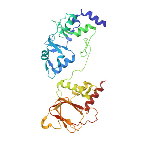
    The family of cystathionine-β-synthase (CBS)-pair domain divalent metal cation transport mediators (CNNMs) is composed of four integral membrane proteins associated with Mg 2+ transport. Structurally, CNNMs contain large cytosolic regions composed of a CBS-pair and a cyclic nucleotide-binding homology (CNBH) domain. How these regulate Mg 2+ transport activity is unknown. Here, we determined the crystal structures of cytosolic fragments in two conformations: Mg 2+ -ATP-analog bound and ligand free. The structures reveal open and closed conformations with functionally important contacts not observed in structures of the individual domains. We also identified a second Mg 2+ -binding region in the CBS-pair domain and a different dimerization interface for the CNBH domain. Analytical ultracentrifugation and isothermal titration calorimetry experiments revealed a tight correlation between Mg 2+ -ATP binding and protein dimerization. Mutations that blocked either function prevented cellular Mg 2+ efflux activity. The results suggest Mg 2+ efflux is regulated by conformational changes associated with Mg 2+ -ATP binding to CNNM CBS-pair domains.


  • Organizational Affiliation
    • Department of Biochemistry & Centre for Structural Biology, McGill University, Montreal, QC H3G 0B1, Canada.

Macromolecule Content 

  • Total Structure Weight: 165.43 kDa 
  • Atom Count: 9,415 
  • Modeled Residue Count: 1,226 
  • Deposited Residue Count: 1,416 
  • Unique protein chains: 1

Macromolecules

Find similar proteins by:|  3D Structure
Entity ID: 1
MoleculeChains  Sequence LengthOrganismDetailsImage
Metal transporter CNNM2,Metal transporter CNNM2
A, B, C, D
354Homo sapiensMutation(s): 0 
Gene Names: CNNM2ACDP2
UniProt & NIH Common Fund Data Resources
Find proteins for Q9H8M5 (Homo sapiens)
Explore Q9H8M5 
Go to UniProtKB:  Q9H8M5
PHAROS:  Q9H8M5
GTEx:  ENSG00000148842 
Entity Groups
Sequence Clusters30% Identity50% Identity70% Identity90% Identity95% Identity100% Identity
UniProt GroupQ9H8M5
Sequence Annotations
Expand
Reference Sequence

Small Molecules

Ligands 2 Unique
IDChains Name / Formula / InChI Key2D Diagram3D Interactions
ANP

Query on ANP


Download:Ideal Coordinates CCD File
E [auth A],
H [auth B],
L [auth C],
N [auth D]
PHOSPHOAMINOPHOSPHONIC ACID-ADENYLATE ESTER
C10 H17 N6 O12 P3
PVKSNHVPLWYQGJ-KQYNXXCUSA-N
MG

Query on MG


Download:Ideal Coordinates CCD File
F [auth A]
G [auth A]
I [auth B]
J [auth B]
K [auth B]
F [auth A],
G [auth A],
I [auth B],
J [auth B],
K [auth B],
M [auth C],
O [auth D],
P [auth D]
MAGNESIUM ION
Mg
JLVVSXFLKOJNIY-UHFFFAOYSA-N

Experimental Data & Validation

Experimental Data

  • Method: X-RAY DIFFRACTION
  • Resolution: 3.50 Å
  • R-Value Free:  0.282 (Depositor), 0.281 (DCC) 
  • R-Value Work:  0.282 (Depositor), 0.241 (DCC) 
Space Group: C 1 2 1
Unit Cell:
Length ( Å )Angle ( ˚ )
a = 290.582α = 90
b = 111.139β = 101.01
c = 103.519γ = 90
Software Package:
Software NamePurpose
PHENIXrefinement
HKL-2000data reduction
HKL-2000data scaling
PHENIXphasing

Structure Validation

View Full Validation Report


Ligand Structure Quality Assessment 


Entry History 

Deposition Data

Revision History  (Full details and data files)

  • Version 1.0: 2019-12-04
    Type: Initial release
  • Version 1.1: 2020-01-01
    Changes: Database references
  • Version 1.2: 2020-03-18
    Changes: Database references
  • Version 1.3: 2023-10-11
    Changes: Data collection, Database references, Derived calculations, Refinement description