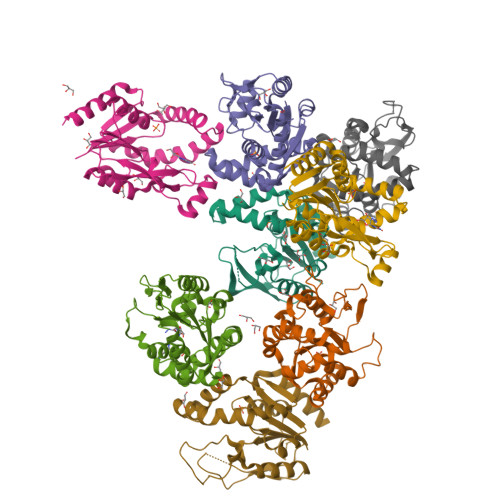
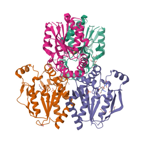
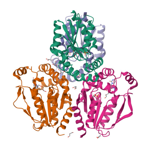
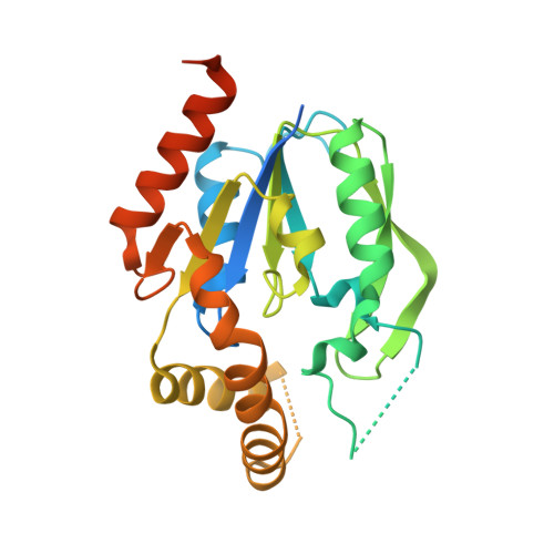
6N55

Crystal structure of human uridine-cytidine kinase 2 complexed with 2'-azidouridine


Experimental Data Snapshot

  • Method: X-RAY DIFFRACTION
  • Resolution: 3.08 Å
  • R-Value Free: 0.258 
  • R-Value Work: 0.211 
  • R-Value Observed: 0.214 

Starting Model: experimental
View more details

wwPDB Validation   3D Report Full Report


Ligand Structure Quality Assessment 


This is version 1.4 of the entry. See complete history


Literature

An optimized chemical-genetic method for cell-specific metabolic labeling of RNA.

Nainar, S.Cuthbert, B.J.Lim, N.M.England, W.E.Ke, K.Sophal, K.Quechol, R.Mobley, D.L.Goulding, C.W.Spitale, R.C.

(2020) Nat Methods 17: 311-318

  • DOI: https://doi.org/10.1038/s41592-019-0726-y
  • Primary Citation of Related Structures:  
    6N53, 6N54, 6N55

  • PubMed Abstract: 

    Tissues and organs are composed of diverse cell types, which poses a major challenge for cell-type-specific profiling of gene expression. Current metabolic labeling methods rely on exogenous pyrimidine analogs that are only incorporated into RNA in cells expressing an exogenous enzyme. This approach assumes that off-target cells cannot incorporate these analogs. We disprove this assumption and identify and characterize the enzymatic pathways responsible for high background incorporation. We demonstrate that mammalian cells can incorporate uracil analogs and characterize the enzymatic pathways responsible for high background incorporation. To overcome these limitations, we developed a new small molecule-enzyme pair consisting of uridine/cytidine kinase 2 and 2'-azidouridine. We demonstrate that 2'-azidouridine is only incorporated in cells expressing uridine/cytidine kinase 2 and characterize selectivity mechanisms using molecular dynamics and X-ray crystallography. Furthermore, this pair can be used to purify and track RNA from specific cellular populations, making it ideal for high-resolution cell-specific RNA labeling. Overall, these results reveal new aspects of mammalian salvage pathways and serve as a new benchmark for designing, characterizing and evaluating methodologies for cell-specific labeling of biomolecules.


  • Organizational Affiliation

    Department of Pharmaceutical Sciences, University of California, Irvine, Irvine, CA, USA.


Macromolecules
Find similar proteins by:  (by identity cutoff)  |  3D Structure
Entity ID: 1
MoleculeChains Sequence LengthOrganismDetailsImage
Uridine-cytidine kinase 2
A, B, C, D, E
A, B, C, D, E, F, G, H
255Homo sapiensMutation(s): 0 
Gene Names: UCK2UMPK
EC: 2.7.1.48
UniProt & NIH Common Fund Data Resources
Find proteins for Q9BZX2 (Homo sapiens)
Explore Q9BZX2 
Go to UniProtKB:  Q9BZX2
PHAROS:  Q9BZX2
GTEx:  ENSG00000143179 
Entity Groups  
Sequence Clusters30% Identity50% Identity70% Identity90% Identity95% Identity100% Identity
UniProt GroupQ9BZX2
Sequence Annotations
Expand
  • Reference Sequence
Small Molecules
Ligands 3 Unique
IDChains Name / Formula / InChI Key2D Diagram3D Interactions
UZ0
Query on UZ0

Download Ideal Coordinates CCD File 
DA [auth D],
JA [auth E],
QA [auth F]
2'-azido-2'-deoxyuridine
C9 H11 N5 O5
MRUKYOQQKHNMFI-XVFCMESISA-N
PO4
Query on PO4

Download Ideal Coordinates CCD File 
EA [auth E]
I [auth A]
KA [auth F]
N [auth B]
R [auth C]
EA [auth E],
I [auth A],
KA [auth F],
N [auth B],
R [auth C],
RA [auth G],
TA [auth H],
W [auth D]
PHOSPHATE ION
O4 P
NBIIXXVUZAFLBC-UHFFFAOYSA-K
GOL
Query on GOL

Download Ideal Coordinates CCD File 
AA [auth D]
BA [auth D]
CA [auth D]
FA [auth E]
GA [auth E]
AA [auth D],
BA [auth D],
CA [auth D],
FA [auth E],
GA [auth E],
HA [auth E],
IA [auth E],
J [auth A],
K [auth A],
L [auth A],
LA [auth F],
M [auth A],
MA [auth F],
NA [auth F],
O [auth B],
OA [auth F],
P [auth B],
PA [auth F],
Q [auth B],
S [auth C],
SA [auth G],
T [auth C],
U [auth C],
V [auth C],
X [auth D],
Y [auth D],
Z [auth D]
GLYCEROL
C3 H8 O3
PEDCQBHIVMGVHV-UHFFFAOYSA-N
Experimental Data & Validation

Experimental Data

  • Method: X-RAY DIFFRACTION
  • Resolution: 3.08 Å
  • R-Value Free: 0.258 
  • R-Value Work: 0.211 
  • R-Value Observed: 0.214 
  • Space Group: P 1 21 1
Unit Cell:
Length ( Å )Angle ( ˚ )
a = 92.747α = 90
b = 85.764β = 95.39
c = 153.367γ = 90
Software Package:
Software NamePurpose
PHENIXrefinement
HKL-2000data reduction
HKL-2000data scaling
PHENIXphasing

Structure Validation

View Full Validation Report


Ligand Structure Quality Assessment 


Entry History & Funding Information

Deposition Data


Funding OrganizationLocationGrant Number
National Institutes of Health/National Institute of Mental Health (NIH/NIMH)United States1R21MH113062

Revision History  (Full details and data files)

  • Version 1.0: 2019-11-27
    Type: Initial release
  • Version 1.1: 2020-01-15
    Changes: Database references
  • Version 1.2: 2020-02-19
    Changes: Database references
  • Version 1.3: 2020-03-18
    Changes: Database references
  • Version 1.4: 2023-10-11
    Changes: Data collection, Database references, Refinement description