6N2D | pdb_00006n2d

Bacillus PS3 ATP synthase membrane region


Experimental Data Snapshot

  • Method: ELECTRON MICROSCOPY
  • Resolution: 3.30 Å
  • Aggregation State: PARTICLE 
  • Reconstruction Method: SINGLE PARTICLE 

wwPDB Validation 3D Report Full Report

Validation slider image for 6N2D

This is version 1.2 of the entry. See complete history

Literature

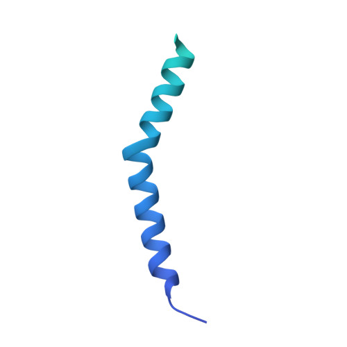
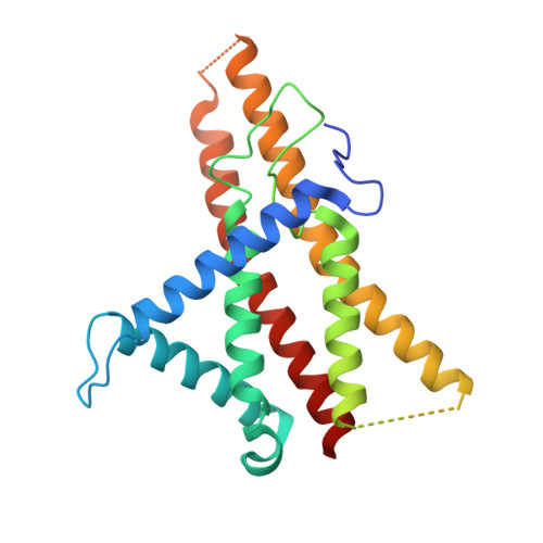
Structure of a bacterial ATP synthase.

Guo, H.Suzuki, T.Rubinstein, J.L.

(2019) Elife 8

  • DOI: https://doi.org/10.7554/eLife.43128
  • Primary Citation Related Structures: 
    6N2D, 6N2Y, 6N2Z, 6N30

  • PubMed Abstract: 

    ATP synthases produce ATP from ADP and inorganic phosphate with energy from a transmembrane proton motive force. Bacterial ATP synthases have been studied extensively because they are the simplest form of the enzyme and because of the relative ease of genetic manipulation of these complexes. We expressed the Bacillus PS3 ATP synthase in Eschericia coli , purified it, and imaged it by cryo-EM, allowing us to build atomic models of the complex in three rotational states. The position of subunit ε shows how it is able to inhibit ATP hydrolysis while allowing ATP synthesis. The architecture of the membrane region shows how the simple bacterial ATP synthase is able to perform the same core functions as the equivalent, but more complicated, mitochondrial complex. The structures reveal the path of transmembrane proton translocation and provide a model for understanding decades of biochemical analysis interrogating the roles of specific residues in the enzyme.


  • Organizational Affiliation
    • The Hospital for Sick Children Research Institute, Toronto, Canada.

Macromolecule Content 

  • Total Structure Weight: 138.33 kDa 
  • Atom Count: 7,162 
  • Modeled Residue Count: 993 
  • Deposited Residue Count: 1,291 
  • Unique protein chains: 3

Macromolecules

Find similar proteins by:|  3D Structure
Entity ID: 1
MoleculeChains  Sequence LengthOrganismDetailsImage
ATP synthase subunit bA [auth b2],
B [auth b1]
167Bacillus sp. PS3Mutation(s): 0 
Gene Names: atpF
Find similar proteins by:|  3D Structure
Entity ID: 2
MoleculeChains  Sequence LengthOrganismDetailsImage
ATP synthase subunit aC [auth a]237Bacillus sp. PS3Mutation(s): 0 
Gene Names: atpB
Find similar proteins by:|  3D Structure
Entity ID: 3
MoleculeChains  Sequence LengthOrganismDetailsImage
ATP synthase subunit c72Bacillus sp. PS3Mutation(s): 0 
Gene Names: atpEuncE
Membrane Entity: Yes 
UniProt
Find proteins for P00845 (Bacillus sp. (strain PS3))
Explore P00845 
Go to UniProtKB:  P00845
Entity Groups
Sequence Clusters30% Identity50% Identity70% Identity90% Identity95% Identity100% Identity
UniProt GroupP00845
Sequence Annotations
Expand
Reference Sequence

Experimental Data & Validation

Experimental Data

  • Method: ELECTRON MICROSCOPY
  • Resolution: 3.30 Å
  • Aggregation State: PARTICLE 
  • Reconstruction Method: SINGLE PARTICLE 
EM Software:
TaskSoftware PackageVersion
RECONSTRUCTIONRELION2.1
MODEL REFINEMENTPHENIX1.14

Structure Validation

View Full Validation Report


Entry History 

& Funding Information

Deposition Data


Funding OrganizationLocationGrant Number
Canadian Institutes of Health Research (CIHR)CanadaMOP 81294
Japan Society for the Promotion of Science (JSPS)JapanJP18H02409

Revision History  (Full details and data files)

  • Version 1.0: 2019-02-20
    Type: Initial release
  • Version 1.1: 2019-12-18
    Changes: Author supporting evidence, Other
  • Version 1.2: 2024-03-20
    Changes: Data collection, Database references