6N04 | pdb_00006n04

The X-ray crystal structure of AbsH3, an FAD dependent reductase from the Abyssomicin biosynthesis pathway in Streptomyces


Experimental Data Snapshot

  • Method: X-RAY DIFFRACTION
  • Resolution: 2.00 Å
  • R-Value Free: 
    0.236 (Depositor), 0.238 (DCC) 
  • R-Value Work: 
    0.183 (Depositor), 0.185 (DCC) 
  • R-Value Observed: 
    0.185 (Depositor) 

Starting Model: experimental
View more details

wwPDB Validation 3D Report Full Report

Validation slider image for 6N04

Ligand Structure Quality Assessment 


This is version 1.3 of the entry. See complete history

Literature

The crystal structure of AbsH3: A putative flavin adenine dinucleotide-dependent reductase in the abyssomicin biosynthesis pathway.

Clinger, J.A.Wang, X.Cai, W.Zhu, Y.Miller, M.D.Zhan, C.G.Van Lanen, S.G.Thorson, J.S.Phillips Jr., G.N.

(2020) Proteins 

  • DOI: https://doi.org/10.1002/prot.25994
  • Primary Citation Related Structures: 
    6N04

  • PubMed Abstract: 

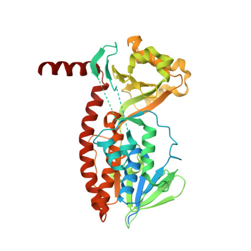
    Natural products and natural product-derived compounds have been widely used for pharmaceuticals for many years, and the search for new natural products that may have interesting activity is ongoing. Abyssomicins are natural product molecules that have antibiotic activity via inhibition of the folate synthesis pathway in microbiota. These compounds also appear to undergo a required [4 + 2] cycloaddition in their biosynthetic pathway. Here we report the structure of an flavin adenine dinucleotide-dependent reductase, AbsH3, from the biosynthetic gene cluster of novel abyssomicins found in Streptomyces sp. LC-6-2.


  • Organizational Affiliation
    • Department of Biosciences, Rice University, Houston, Texas, USA.

Macromolecule Content 

  • Total Structure Weight: 93.82 kDa 
  • Atom Count: 6,559 
  • Modeled Residue Count: 731 
  • Deposited Residue Count: 868 
  • Unique protein chains: 1

Macromolecules

Find similar proteins by:|  3D Structure
Entity ID: 1
MoleculeChains  Sequence LengthOrganismDetailsImage
AbsH3
A, B
434Streptomyces sp. LC-6-2Mutation(s): 0 
EC: 1.14.13
UniProt
Find proteins for A0A1V0QH64 (Streptomyces sp. LC-6-2)
Explore A0A1V0QH64 
Go to UniProtKB:  A0A1V0QH64
Entity Groups
Sequence Clusters30% Identity50% Identity70% Identity90% Identity95% Identity100% Identity
UniProt GroupA0A1V0QH64
Sequence Annotations
Expand
Reference Sequence

Experimental Data & Validation

Experimental Data

  • Method: X-RAY DIFFRACTION
  • Resolution: 2.00 Å
  • R-Value Free:  0.236 (Depositor), 0.238 (DCC) 
  • R-Value Work:  0.183 (Depositor), 0.185 (DCC) 
  • R-Value Observed: 0.185 (Depositor) 
Space Group: P 21 21 21
Unit Cell:
Length ( Å )Angle ( ˚ )
a = 53.316α = 90
b = 109.491β = 90
c = 143.637γ = 90
Software Package:
Software NamePurpose
PHENIXrefinement
XDSdata reduction
XSCALEdata scaling
PHASERphasing
PDB_EXTRACTdata extraction

Structure Validation

View Full Validation Report


Ligand Structure Quality Assessment 


Entry History 

& Funding Information

Deposition Data


Funding OrganizationLocationGrant Number
National Institutes of Health/National Institute of General Medical Sciences (NIH/NIGMS)United StatesR01 CA217255-01A1
National Institutes of Health/National Institute of General Medical Sciences (NIH/NIGMS)United StatesR01 GM115261-02

Revision History  (Full details and data files)

  • Version 1.0: 2019-11-13
    Type: Initial release
  • Version 1.1: 2020-01-01
    Changes: Author supporting evidence
  • Version 1.2: 2022-02-23
    Changes: Database references
  • Version 1.3: 2023-10-11
    Changes: Data collection, Refinement description