6MX4 | pdb_00006mx4

CryoEM structure of chimeric Eastern Equine Encephalitis Virus


Experimental Data Snapshot

  • Method: ELECTRON MICROSCOPY
  • Resolution: 4.40 Å
  • Aggregation State: PARTICLE 
  • Reconstruction Method: SINGLE PARTICLE 

wwPDB Validation 3D Report Full Report

Validation slider image for 6MX4

This is version 2.1 of the entry. See complete history. 

Literature

Cryo-EM Structures of Eastern Equine Encephalitis Virus Reveal Mechanisms of Virus Disassembly and Antibody Neutralization.

Hasan, S.S., Sun, C., Kim, A.S., Watanabe, Y., Chen, C.L., Klose, T., Buda, G., Crispin, M., Diamond, M.S., Klimstra, W.B., Rossmann, M.G.

(2018) Cell Rep 25: 3136-3147.e5

  • DOI: https://doi.org/10.1016/j.celrep.2018.11.067
  • Primary Citation Related Structures: 
    6MUI, 6MW9, 6MWC, 6MWV, 6MWX, 6MX4, 6MX7

  • PubMed Abstract: 

    Alphaviruses are enveloped pathogens that cause arthritis and encephalitis. Here, we report a 4.4-Å cryoelectron microscopy (cryo-EM) structure of eastern equine encephalitis virus (EEEV), an alphavirus that causes fatal encephalitis in humans. Our analysis provides insights into viral entry into host cells. The envelope protein E2 showed a binding site for the cellular attachment factor heparan sulfate. The presence of a cryptic E2 glycan suggests how EEEV escapes surveillance by lectin-expressing myeloid lineage cells, which are sentinels of the immune system. A mechanism for nucleocapsid core release and disassembly upon viral entry was inferred based on pH changes and capsid dissociation from envelope proteins. The EEEV capsid structure showed a viral RNA genome binding site adjacent to a ribosome binding site for viral genome translation following genome release. Using five Fab-EEEV complexes derived from neutralizing antibodies, our investigation provides insights into EEEV host cell interactions and protective epitopes relevant to vaccine design.


  • Organizational Affiliation: 
    • Department of Biological Sciences, Purdue University, West Lafayette, IN 47907, USA.

Macromolecule Content 

  • Total Structure Weight: 498.78 kDa 
  • Atom Count: 31,491 
  • Modeled Residue Count: 4,039 
  • Deposited Residue Count: 4,484 
  • Unique protein chains: 3

Macromolecules

Find similar proteins by:|  3D Structure
Entity ID: 1
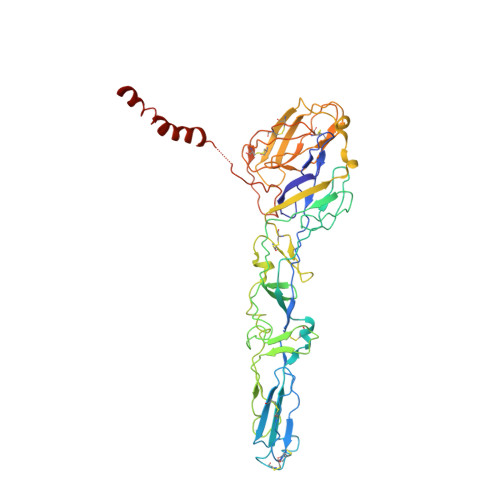
MoleculeChains  Sequence LengthOrganismDetailsImage
E1
A, D, G, J
441Eastern equine encephalitis virusMutation(s): 0 
Membrane Entity: Yes 
UniProt
Find proteins for Q4QXJ7 (Eastern equine encephalitis virus (strain Florida 91-469))
Explore Q4QXJ7 
Go to UniProtKB:  Q4QXJ7
Entity Groups
Sequence Clusters30% Identity50% Identity70% Identity90% Identity95% Identity100% Identity
UniProt GroupQ4QXJ7
Glycosylation
Glycosylation Sites: 1
Sequence Annotations
Expand
Reference Sequence
Find similar proteins by:|  3D Structure
Entity ID: 2
MoleculeChains  Sequence LengthOrganismDetailsImage
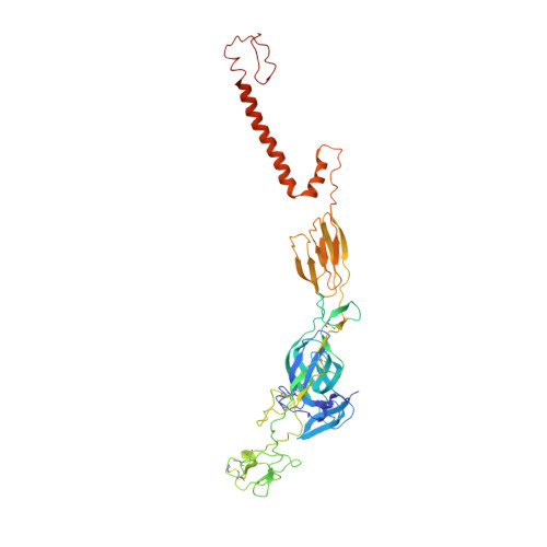
E2
B, E, H, K
420Eastern equine encephalitis virusMutation(s): 0 
Membrane Entity: Yes 
UniProt
Find proteins for Q4QXJ7 (Eastern equine encephalitis virus (strain Florida 91-469))
Explore Q4QXJ7 
Go to UniProtKB:  Q4QXJ7
Entity Groups
Sequence Clusters30% Identity50% Identity70% Identity90% Identity95% Identity100% Identity
UniProt GroupQ4QXJ7
Glycosylation
Glycosylation Sites: 1
Sequence Annotations
Expand
Reference Sequence
Find similar proteins by:|  3D Structure
Entity ID: 3
MoleculeChains  Sequence LengthOrganismDetailsImage
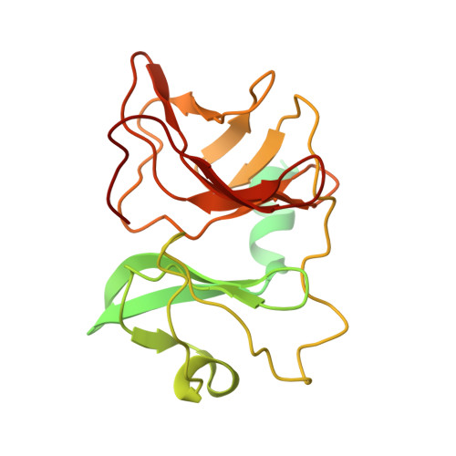
Capsid
C, F, I, L
260Eastern equine encephalitis virusMutation(s): 0 
Membrane Entity: Yes 
UniProt
Find proteins for P27284 (Eastern equine encephalitis virus (strain va33[ten broeck]))
Explore P27284 
Go to UniProtKB:  P27284
Entity Groups
Sequence Clusters30% Identity50% Identity70% Identity90% Identity95% Identity100% Identity
UniProt GroupP27284
Sequence Annotations
Expand
Reference Sequence

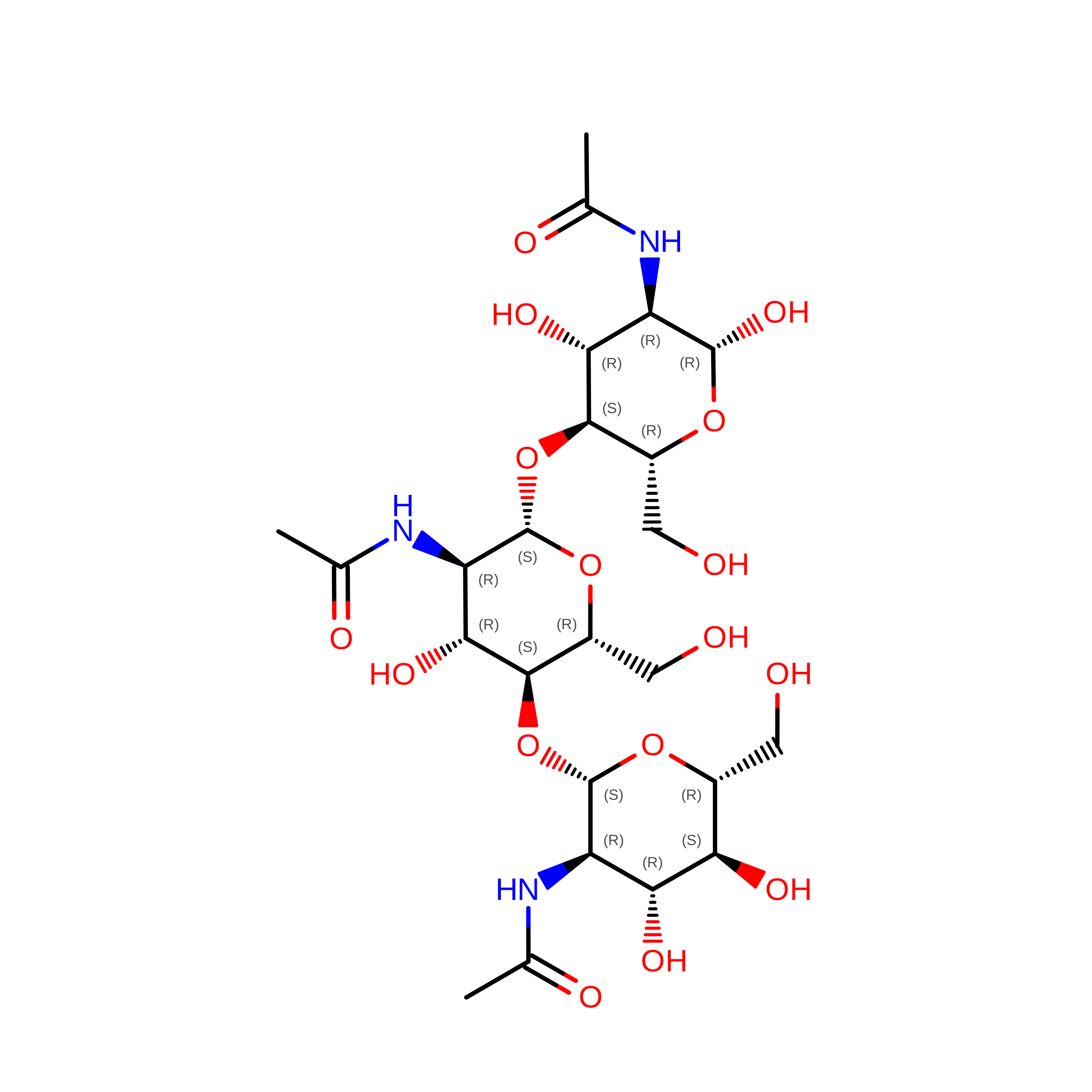
Oligosaccharides

Help  
Entity ID: 4
MoleculeChains Length2D Diagram GlycosylationD Interactions
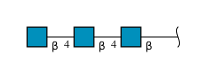
2-acetamido-2-deoxy-beta-D-glucopyranose-(1-4)-2-acetamido-2-deoxy-beta-D-glucopyranose-(1-4)-2-acetamido-2-deoxy-beta-D-glucopyranose
M, N, O, P
3N-Glycosylation
Glycosylation Resources
GlyTouCan: G47362BJ
GlyCosmos: G47362BJ
GlyGen: G47362BJ

Experimental Data & Validation

Experimental Data

  • Method: ELECTRON MICROSCOPY
  • Resolution: 4.40 Å
  • Aggregation State: PARTICLE 
  • Reconstruction Method: SINGLE PARTICLE 

Structure Validation

View Full Validation Report


Entry History 

& Funding Information

Deposition Data


Funding OrganizationLocationGrant Number
National Institutes of Health/National Institute Of Allergy and Infectious Diseases (NIH/NIAID)United StatesR01AI095366
National Institutes of Health/National Institute Of Allergy and Infectious Diseases (NIH/NIAID)United States2R01AI095436
National Institutes of Health/National Institute Of Allergy and Infectious Diseases (NIH/NIAID)United StatesR21AI117331-01
Bill & Melinda Gates FoundationUnited KingdomOPP1115782

Revision History  (Full details and data files)

  • Version 1.0: 2018-12-19
    Type: Initial release
  • Version 1.1: 2018-12-26
    Changes: Data collection, Database references
  • Version 1.2: 2019-12-18
    Changes: Author supporting evidence
  • Version 2.0: 2020-07-29
    Type: Remediation
    Reason: Carbohydrate remediation
    Changes: Atomic model, Data collection, Derived calculations, Structure summary
  • Version 2.1: 2024-10-23
    Changes: Data collection, Database references, Derived calculations, Structure summary