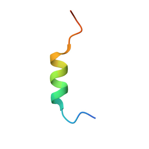
6MV5

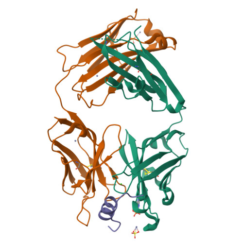
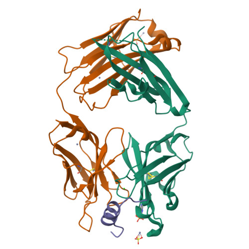
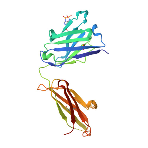
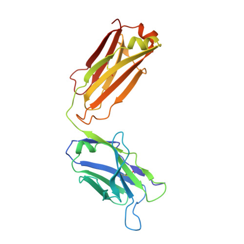
Anti-PCSK9 fab 6E2 bound to the N-terminal peptide from PCSK9 (E32K)


Experimental Data Snapshot

  • Method: X-RAY DIFFRACTION
  • Resolution: 2.10 Å
  • R-Value Free: 0.238 
  • R-Value Work: 0.210 
  • R-Value Observed: 0.211 

Starting Model: experimental
View more details

wwPDB Validation   3D Report Full Report


This is version 1.1 of the entry. See complete history


Literature

Identification of a Helical Segment within the Intrinsically Disordered Region of the PCSK9 Prodomain.

Ultsch, M.Li, W.Eigenbrot, C.Di Lello, P.Lipari, M.T.Gerhardy, S.AhYoung, A.P.Quinn, J.Franke, Y.Chen, Y.Kong Beltran, M.Peterson, A.Kirchhofer, D.

(2019) J Mol Biol 431: 885-903

  • DOI: https://doi.org/10.1016/j.jmb.2018.11.025
  • Primary Citation of Related Structures:  
    6E4Y, 6E4Z, 6MV5

  • PubMed Abstract: 

    Proprotein convertase subtilisin/kexin 9 (PCSK9) is a key regulator of lipid metabolism by degrading liver LDL receptors. Structural studies have provided molecular details of PCSK9 function. However, the N-terminal acidic stretch of the PCSK9 prodomain (Q31-T60) has eluded structural investigation, since it is in a disordered state. The interest in this region is intensified by the presence of human missense mutations associated with low and high LDL-c levels (E32K, D35Y, and R46L, respectively), as well as two posttranslationally modified sites, sulfated Y38 and phosphorylated S47. Herein we show that a segment within this region undergoes disorder-to-order transition. Experiments with acidic stretch-derived peptides demonstrated that the folding is centered at the segment Y38-L45, which adopts an α-helix as determined by NMR analysis of free peptides and by X-ray crystallography of peptides in complex with antibody 6E2 (Ab6E2). In the Fab6E2-peptide complexes, the structured region features a central 2 1/4-turn α-helix and encompasses up to 2/3 of the length of the acidic stretch, including the missense mutations and posttranslationally modified sites. Experiments with helix-breaking proline substitutions in peptides and in PCSK9 protein indicated that Ab6E2 specifically recognizes the helical conformation of the acidic stretch. Therefore, the observed quantitative binding of Ab6E2 to native PCSK9 from various cell lines suggests that the disorder-to-order transition is a true feature of PCSK9 and not limited to peptides. Because the helix provides a constrained spatial orientation of the missense mutations and the posttranslationally modified residues, it is probable that their biological functions take place in the context of an ordered conformational state.


  • Organizational Affiliation

    Department of Structural Biology, Genentech, Inc., 1 DNA Way, South San Francisco, CA 94080, USA.


Macromolecules
Find similar proteins by:  (by identity cutoff)  |  3D Structure
Entity ID: 1
MoleculeChains Sequence LengthOrganismDetailsImage
Anti-PCSK9 fab 6E2 heavy chainA [auth H]224Mus musculusMutation(s): 0 
Entity Groups  
Sequence Clusters30% Identity50% Identity70% Identity90% Identity95% Identity100% Identity
Sequence Annotations
Expand
  • Reference Sequence
Find similar proteins by:  (by identity cutoff)  |  3D Structure
Entity ID: 2
MoleculeChains Sequence LengthOrganismDetailsImage
Anti-PCSK9 fab 6E2 light chainB [auth L]219Mus musculusMutation(s): 0 
UniProt
Find proteins for A0A097PUG4 (Mus musculus)
Explore A0A097PUG4 
Go to UniProtKB:  A0A097PUG4
Entity Groups  
Sequence Clusters30% Identity50% Identity70% Identity90% Identity95% Identity100% Identity
UniProt GroupA0A097PUG4
Sequence Annotations
Expand
  • Reference Sequence

Find similar proteins by:  Sequence   |   3D Structure  

Entity ID: 3
MoleculeChains Sequence LengthOrganismDetailsImage
Proprotein convertase subtilisin/kexin type 9C [auth P]23Homo sapiensMutation(s): 2 
EC: 3.4.21
UniProt & NIH Common Fund Data Resources
Find proteins for Q8NBP7 (Homo sapiens)
Explore Q8NBP7 
Go to UniProtKB:  Q8NBP7
PHAROS:  Q8NBP7
GTEx:  ENSG00000169174 
Entity Groups  
Sequence Clusters30% Identity50% Identity70% Identity90% Identity95% Identity100% Identity
UniProt GroupQ8NBP7
Sequence Annotations
Expand
  • Reference Sequence
Small Molecules
Ligands 3 Unique
IDChains Name / Formula / InChI Key2D Diagram3D Interactions
SO4
Query on SO4

Download Ideal Coordinates CCD File 
F [auth H],
J [auth L]
SULFATE ION
O4 S
QAOWNCQODCNURD-UHFFFAOYSA-L
ZN
Query on ZN

Download Ideal Coordinates CCD File 
D [auth H],
G [auth L],
H [auth L],
I [auth L]
ZINC ION
Zn
PTFCDOFLOPIGGS-UHFFFAOYSA-N
CL
Query on CL

Download Ideal Coordinates CCD File 
E [auth H]CHLORIDE ION
Cl
VEXZGXHMUGYJMC-UHFFFAOYSA-M
Modified Residues  1 Unique
IDChains TypeFormula2D DiagramParent
NEP
Query on NEP
A [auth H]L-PEPTIDE LINKINGC6 H10 N3 O5 PHIS
Experimental Data & Validation

Experimental Data

  • Method: X-RAY DIFFRACTION
  • Resolution: 2.10 Å
  • R-Value Free: 0.238 
  • R-Value Work: 0.210 
  • R-Value Observed: 0.211 
  • Space Group: P 21 21 2
Unit Cell:
Length ( Å )Angle ( ˚ )
a = 70.449α = 90
b = 100.42β = 90
c = 86.59γ = 90
Software Package:
Software NamePurpose
PHENIXrefinement
MOSFLMdata reduction
XDSdata scaling
PHASERphasing

Structure Validation

View Full Validation Report


Entry History 

Deposition Data

Revision History  (Full details and data files)

  • Version 1.0: 2019-04-10
    Type: Initial release
  • Version 1.1: 2023-10-11
    Changes: Data collection, Database references, Derived calculations, Refinement description