


Crystal Structure of Prolyl-tRNA Synthetase from Onchocerca volvulus with bound Halofuginone and nucleotide
Dranow, D.M., Conrady, D.G., Lorimer, D.D., Horanyi, P.S., Edwards, T.E.To be published.
Experimental Data Snapshot
Starting Model: experimental
View more details
Entity ID: 1 | |||||
|---|---|---|---|---|---|
| Molecule | Chains | Sequence Length | Organism | Details | Image |
| Uncharacterized protein | 505 | Onchocerca volvulus | Mutation(s): 0 EC: 6.1.1.15 | 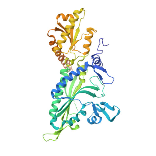 | |
UniProt | |||||
Entity Groups | |||||
| Sequence Clusters | 30% Identity50% Identity70% Identity90% Identity95% Identity100% Identity | ||||
| UniProt Group | A0A2K6VKP7 | ||||
Sequence AnnotationsExpand | |||||
Reference Sequence | |||||
| Ligands 7 Unique | |||||
|---|---|---|---|---|---|
| ID | Chains | Name / Formula / InChI Key | 2D Diagram | 3D Interactions | |
| ANP (Subject of Investigation/LOI) Download:Ideal Coordinates CCD File | E [auth A] | PHOSPHOAMINOPHOSPHONIC ACID-ADENYLATE ESTER C10 H17 N6 O12 P3 PVKSNHVPLWYQGJ-KQYNXXCUSA-N |  | ||
| HFG (Subject of Investigation/LOI) Download:Ideal Coordinates CCD File | D [auth A] | 7-bromo-6-chloro-3-{3-[(2R,3S)-3-hydroxypiperidin-2-yl]-2-oxopropyl}quinazolin-4(3H)-one C16 H17 Br Cl N3 O3 LVASCWIMLIKXLA-CABCVRRESA-N |  | ||
| AMP (Subject of Investigation/LOI) Download:Ideal Coordinates CCD File | F [auth A] | ADENOSINE MONOPHOSPHATE C10 H14 N5 O7 P UDMBCSSLTHHNCD-KQYNXXCUSA-N |  | ||
| 2PN (Subject of Investigation/LOI) Download:Ideal Coordinates CCD File | G [auth A] | IMIDODIPHOSPHORIC ACID H5 N O6 P2 GNGSOPFGGKKDQP-UHFFFAOYSA-N |  | ||
| SER Download:Ideal Coordinates CCD File | K [auth A], L [auth A] | SERINE C3 H7 N O3 MTCFGRXMJLQNBG-REOHCLBHSA-N |  | ||
| GLY Download:Ideal Coordinates CCD File | H [auth A], I [auth A], J [auth A] | GLYCINE C2 H5 N O2 DHMQDGOQFOQNFH-UHFFFAOYSA-N |  | ||
| MG Download:Ideal Coordinates CCD File | B [auth A], C [auth A] | MAGNESIUM ION Mg JLVVSXFLKOJNIY-UHFFFAOYSA-N |  | ||
| Length ( Å ) | Angle ( ˚ ) |
|---|---|
| a = 159.72 | α = 90 |
| b = 159.72 | β = 90 |
| c = 143.72 | γ = 120 |
| Software Name | Purpose |
|---|---|
| PHENIX | refinement |
| XDS | data reduction |
| XSCALE | data scaling |
| PDB_EXTRACT | data extraction |
| MoRDa | phasing |


