6MLK

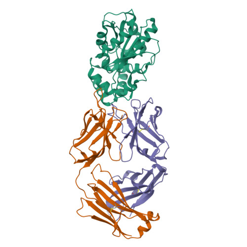
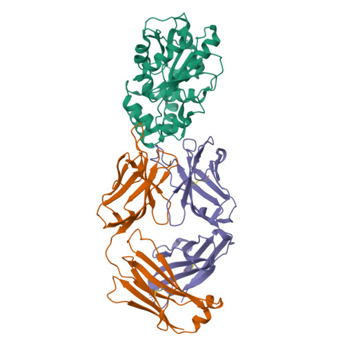
Structure of Thioesterase from DEBS with a thioesterase-specific antibody


Experimental Data Snapshot

  • Method: X-RAY DIFFRACTION
  • Resolution: 2.45 Å
  • R-Value Free: 0.235 
  • R-Value Work: 0.175 
  • R-Value Observed: 0.178 

Starting Models: experimental
View more details

wwPDB Validation   3D Report Full Report


This is version 1.4 of the entry. See complete history


Literature

Discovery and Characterization of a Thioesterase-Specific Monoclonal Antibody That Recognizes the 6-Deoxyerythronolide B Synthase.

Li, X.Sevillano, N.La Greca, F.Hsu, J.Mathews, I.I.Matsui, T.Craik, C.S.Khosla, C.

(2018) Biochemistry 57: 6201-6208

  • DOI: https://doi.org/10.1021/acs.biochem.8b00886
  • Primary Citation of Related Structures:  
    6MLK

  • PubMed Abstract: 

    Assembly line polyketide synthases (PKSs) are large multimodular enzymes responsible for the biosynthesis of diverse antibiotics in bacteria. Structural and mechanistic analysis of these megasynthases can benefit from the discovery of reagents that recognize individual domains or linkers in a site-specific manner. Monoclonal antibodies not only have proven themselves as premier tools in analogous applications but also have the added benefit of constraining the conformational flexibility of their targets in unpredictable but often useful ways. Here we have exploited a library based on the naïve human antibody repertoire to discover a F ab (3A6) that recognizes the terminal thioesterase (TE) domain of the 6-deoxyerythronolide B synthase with high specificity. Biochemical assays were used to verify that 3A6 binding does not inhibit enzyme turnover. The co-crystal structure of the TE-3A6 complex was determined at 2.45 Å resolution, resulting in atomic characterization of this protein-protein recognition mechanism. F ab binding had minimal effects on the structural integrity of the TE. In turn, these insights were used to interrogate via small-angle X-ray scattering the solution-phase conformation of 3A6 complexed to a catalytically competent PKS module and bimodule. Altogether, we have developed a high-affinity monoclonal antibody tool that recognizes the TE domain of the 6-deoxyerythronolide B synthase while maintaining its native function.


  • Organizational Affiliation

    Department of Pharmaceutical Chemistry , University of California San Francisco , San Francisco , California 94158 , United States.


Macromolecules
Find similar proteins by:  (by identity cutoff)  |  3D Structure
Entity ID: 1
MoleculeChains Sequence LengthOrganismDetailsImage
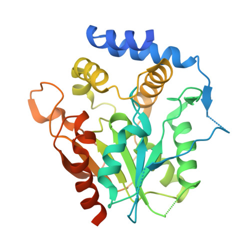
6-deoxyerythronolide-B synthase EryA3, modules 5 and 6300Saccharopolyspora erythraeaMutation(s): 0 
Gene Names: eryA
EC: 2.3.1.94
UniProt
Find proteins for Q03133 (Saccharopolyspora erythraea)
Explore Q03133 
Go to UniProtKB:  Q03133
Entity Groups  
Sequence Clusters30% Identity50% Identity70% Identity90% Identity95% Identity100% Identity
UniProt GroupQ03133
Sequence Annotations
Expand
  • Reference Sequence
Find similar proteins by:  (by identity cutoff)  |  3D Structure
Entity ID: 2
MoleculeChains Sequence LengthOrganismDetailsImage
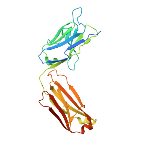
Light chain of Fab 3A6B [auth L]235Homo sapiensMutation(s): 0 
Entity Groups  
Sequence Clusters30% Identity50% Identity70% Identity90% Identity95% Identity100% Identity
Sequence Annotations
Expand
  • Reference Sequence
Find similar proteins by:  (by identity cutoff)  |  3D Structure
Entity ID: 3
MoleculeChains Sequence LengthOrganismDetailsImage
Heavy chain of Fab 3A6C [auth H]251Homo sapiensMutation(s): 0 
Entity Groups  
Sequence Clusters30% Identity50% Identity70% Identity90% Identity95% Identity100% Identity
Sequence Annotations
Expand
  • Reference Sequence
Small Molecules
Ligands 1 Unique
IDChains Name / Formula / InChI Key2D Diagram3D Interactions
CL
Query on CL

Download Ideal Coordinates CCD File 
D [auth H]CHLORIDE ION
Cl
VEXZGXHMUGYJMC-UHFFFAOYSA-M
Experimental Data & Validation

Experimental Data

  • Method: X-RAY DIFFRACTION
  • Resolution: 2.45 Å
  • R-Value Free: 0.235 
  • R-Value Work: 0.175 
  • R-Value Observed: 0.178 
  • Space Group: H 3 2
Unit Cell:
Length ( Å )Angle ( ˚ )
a = 172.58α = 90
b = 172.58β = 90
c = 159.05γ = 120
Software Package:
Software NamePurpose
XSCALEdata scaling
PHENIXrefinement
PDB_EXTRACTdata extraction
XDSdata reduction
MOLREPphasing

Structure Validation

View Full Validation Report


Entry History & Funding Information

Deposition Data


Funding OrganizationLocationGrant Number
National Institutes of Health/National Human Genome Research Institute (NIH/NHGRI)United StatesGM087934
National Institutes of Health/National Human Genome Research Institute (NIH/NHGRI)United StatesP41CA196276
National Institutes of Health/National Human Genome Research Institute (NIH/NHGRI)United StatesGM104659
Department of Energy (DOE, United States)United StatesDE-AC02-76SF00515
Department of Energy (DOE, United States)United StatesP41GM103393

Revision History  (Full details and data files)

  • Version 1.0: 2018-10-17
    Type: Initial release
  • Version 1.1: 2018-11-07
    Changes: Data collection, Database references
  • Version 1.2: 2019-12-04
    Changes: Author supporting evidence
  • Version 1.3: 2023-10-11
    Changes: Data collection, Database references, Refinement description
  • Version 1.4: 2024-10-16
    Changes: Structure summary