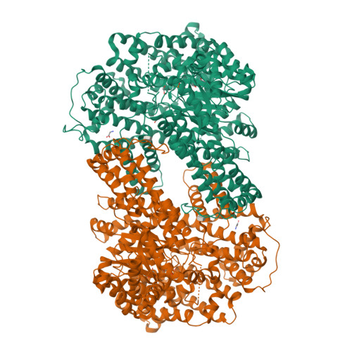
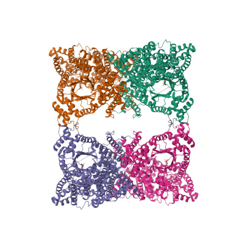
6MGI

Photosynthetic phosphoenolpyruvate carboxylase isoenzyme from maize complexed with the allosteric activator glucose-6-phosphate in its allosteric site


Experimental Data Snapshot

  • Method: X-RAY DIFFRACTION
  • Resolution: 2.99 Å
  • R-Value Free: 0.243 
  • R-Value Work: 0.208 
  • R-Value Observed: 0.210 

Starting Model: experimental
View more details

wwPDB Validation   3D Report Full Report


Ligand Structure Quality Assessment 

Created with Raphaël 2.3.0Worse 01 BetterLigand structure goodness of fit to experimental dataBest fitted G6PClick on this verticalbar to view detailsBest fitted PGAClick on this verticalbar to view details

This is version 1.4 of the entry. See complete history


Literature

Structural and biochemical evidence of the glucose 6-phosphate-allosteric site of maize C4-phosphoenolpyruvate carboxylase: its importance in the overall enzyme kinetics.

Munoz-Clares, R.A.Gonzalez-Segura, L.Juarez-Diaz, J.A.Mujica-Jimenez, C.

(2020) Biochem J 477: 2095-2114

  • DOI: https://doi.org/10.1042/BCJ20200304
  • Primary Citation of Related Structures:  
    6MGI

  • PubMed Abstract: 

    Activation of phosphoenolpyruvate carboxylase (PEPC) enzymes by glucose 6-phosphate (G6P) and other phospho-sugars is of major physiological relevance. Previous kinetic, site-directed mutagenesis and crystallographic results are consistent with allosteric activation, but the existence of a G6P-allosteric site was questioned and competitive activation-in which G6P would bind to the active site eliciting the same positive homotropic effect as the substrate phosphoenolpyruvate (PEP)-was proposed. Here, we report the crystal structure of the PEPC-C4 isozyme from Zea mays with G6P well bound into the previously proposed allosteric site, unambiguously confirming its existence. To test its functionality, Asp239-which participates in a web of interactions of the protein with G6P-was changed to alanine. The D239A variant was not activated by G6P but, on the contrary, inhibited. Inhibition was also observed in the wild-type enzyme at concentrations of G6P higher than those producing activation, and probably arises from G6P binding to the active site in competition with PEP. The lower activity and cooperativity for the substrate PEP, lower activation by glycine and diminished response to malate of the D239A variant suggest that the heterotropic allosteric activation effects of free-PEP are also abolished in this variant. Together, our findings are consistent with both the existence of the G6P-allosteric site and its essentiality for the activation of PEPC enzymes by phosphorylated compounds. Furthermore, our findings suggest a central role of the G6P-allosteric site in the overall kinetics of these enzymes even in the absence of G6P or other phospho-sugars, because of its involvement in activation by free-PEP.


  • Organizational Affiliation

    Departamento de Bioquímica, Facultad de Química, Universidad Nacional Autónoma de México, Ciudad Universitaria, 04510 Ciudad de México, Mexico.


Macromolecules
Find similar proteins by:  (by identity cutoff)  |  3D Structure
Entity ID: 1
MoleculeChains Sequence LengthOrganismDetailsImage
Phosphoenolpyruvate carboxylase
A, B
970Zea maysMutation(s): 0 
Gene Names: ppc1
EC: 4.1.1.31
UniProt
Find proteins for P04711 (Zea mays)
Explore P04711 
Go to UniProtKB:  P04711
Entity Groups  
Sequence Clusters30% Identity50% Identity70% Identity90% Identity95% Identity100% Identity
UniProt GroupP04711
Sequence Annotations
Expand
  • Reference Sequence
Small Molecules
Ligands 5 Unique
IDChains Name / Formula / InChI Key2D Diagram3D Interactions
G6P
Query on G6P

Download Ideal Coordinates CCD File 
D [auth A],
K [auth B]
6-O-phosphono-alpha-D-glucopyranose
C6 H13 O9 P
NBSCHQHZLSJFNQ-DVKNGEFBSA-N
PGA
Query on PGA

Download Ideal Coordinates CCD File 
I [auth A],
P [auth B]
2-PHOSPHOGLYCOLIC ACID
C2 H5 O6 P
ASCFNMCAHFUBCO-UHFFFAOYSA-N
GOL
Query on GOL

Download Ideal Coordinates CCD File 
N [auth B],
O [auth B]
GLYCEROL
C3 H8 O3
PEDCQBHIVMGVHV-UHFFFAOYSA-N
GLY
Query on GLY

Download Ideal Coordinates CCD File 
C [auth A],
J [auth B]
GLYCINE
C2 H5 N O2
DHMQDGOQFOQNFH-UHFFFAOYSA-N
EDO
Query on EDO

Download Ideal Coordinates CCD File 
E [auth A]
F [auth A]
G [auth A]
H [auth A]
L [auth B]
1,2-ETHANEDIOL
C2 H6 O2
LYCAIKOWRPUZTN-UHFFFAOYSA-N
Experimental Data & Validation

Experimental Data

  • Method: X-RAY DIFFRACTION
  • Resolution: 2.99 Å
  • R-Value Free: 0.243 
  • R-Value Work: 0.208 
  • R-Value Observed: 0.210 
  • Space Group: C 2 2 21
Unit Cell:
Length ( Å )Angle ( ˚ )
a = 152.087α = 90
b = 170.679β = 90
c = 244.622γ = 90
Software Package:
Software NamePurpose
PHENIXrefinement
XDSdata reduction
SCALAdata scaling
PHASERphasing

Structure Validation

View Full Validation Report


Ligand Structure Quality Assessment 

Created with Raphaël 2.3.0Worse 01 BetterLigand structure goodness of fit to experimental dataBest fitted G6PClick on this verticalbar to view detailsBest fitted PGAClick on this verticalbar to view details

Entry History & Funding Information

Deposition Data


Funding OrganizationLocationGrant Number
Programa de Apoyo a Proyectos de Investigacion e Innovacion TecnologicaMexicoIN216911

Revision History  (Full details and data files)

  • Version 1.0: 2019-09-18
    Type: Initial release
  • Version 1.1: 2019-10-23
    Changes: Author supporting evidence, Data collection, Derived calculations
  • Version 1.2: 2020-06-24
    Changes: Database references
  • Version 1.3: 2020-07-29
    Type: Remediation
    Reason: Carbohydrate remediation
    Changes: Data collection, Derived calculations, Structure summary
  • Version 1.4: 2023-10-11
    Changes: Data collection, Database references, Refinement description, Structure summary