6MDP | pdb_00006mdp

The D1 and D2 domain rings of NSF engaging the SNAP-25 N-terminus within the 20S supercomplex (focused refinement on D1/D2 rings, class 2)


Experimental Data Snapshot

  • Method: ELECTRON MICROSCOPY
  • Resolution: 3.80 Å
  • Aggregation State: PARTICLE 
  • Reconstruction Method: SINGLE PARTICLE 

Starting Model: experimental
View more details

wwPDB Validation 3D Report Full Report

Validation slider image for 6MDP

This is version 1.3 of the entry. See complete history

Literature

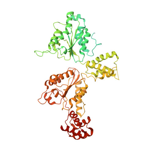
Structural principles of SNARE complex recognition by the AAA+ protein NSF.

White, K.I.Zhao, M.Choi, U.B.Pfuetzner, R.A.Brunger, A.T.

(2018) Elife 7

  • DOI: https://doi.org/10.7554/eLife.38888
  • Primary Citation Related Structures: 
    6MDM, 6MDN, 6MDO, 6MDP

  • PubMed Abstract: 

    The recycling of SNARE proteins following complex formation and membrane fusion is an essential process in eukaryotic trafficking. A highly conserved AAA+ protein, NSF ( N -ethylmaleimide sensitive factor) and an adaptor protein, SNAP (soluble NSF attachment protein), disassemble the SNARE complex. We report electron-cryomicroscopy structures of the complex of NSF, αSNAP, and the full-length soluble neuronal SNARE complex (composed of syntaxin-1A, synaptobrevin-2, SNAP-25A) in the presence of ATP under non-hydrolyzing conditions at ~3.9 Å resolution. These structures reveal electrostatic interactions by which two αSNAP molecules interface with a specific surface of the SNARE complex. This interaction positions the SNAREs such that the 15 N-terminal residues of SNAP-25A are loaded into the D1 ring pore of NSF via a spiral pattern of interactions between a conserved tyrosine NSF residue and SNAP-25A backbone atoms. This loading process likely precedes ATP hydrolysis. Subsequent ATP hydrolysis then drives complete disassembly.


  • Organizational Affiliation
    • Department of Molecular and Cellular Physiology, Stanford University, Stanford, United States.

Macromolecule Content 

  • Total Structure Weight: 541.99 kDa 
  • Atom Count: 22,433 
  • Modeled Residue Count: 2,832 
  • Deposited Residue Count: 4,815 
  • Unique protein chains: 2

Macromolecules

Find similar proteins by:|  3D Structure
Entity ID: 1
MoleculeChains  Sequence LengthOrganismDetailsImage
Vesicle-fusing ATPase
A, B, C, D, E
A, B, C, D, E, F
768Cricetulus griseusMutation(s): 0 
Gene Names: NSF
EC: 3.6.4.6
UniProt
Find proteins for P18708 (Cricetulus griseus)
Explore P18708 
Go to UniProtKB:  P18708
Entity Groups
Sequence Clusters30% Identity50% Identity70% Identity90% Identity95% Identity100% Identity
UniProt GroupP18708
Sequence Annotations
Expand
Reference Sequence
Find similar proteins by:|  3D Structure
Entity ID: 2
MoleculeChains  Sequence LengthOrganismDetailsImage
Synaptosomal-associated protein 25G [auth H]207Rattus norvegicusMutation(s): 0 
Gene Names: Snap25Snap
UniProt
Find proteins for P60881 (Rattus norvegicus)
Explore P60881 
Go to UniProtKB:  P60881
Entity Groups
Sequence Clusters30% Identity50% Identity70% Identity90% Identity95% Identity100% Identity
UniProt GroupP60881
Sequence Annotations
Expand
Reference Sequence

Small Molecules

Ligands 2 Unique
IDChains Name / Formula / InChI Key2D Diagram3D Interactions
ATP

Query on ATP


Download:Ideal Coordinates CCD File
I [auth A]
J [auth B]
K [auth B]
L [auth C]
M [auth C]
I [auth A],
J [auth B],
K [auth B],
L [auth C],
M [auth C],
N [auth D],
O [auth D],
P [auth E],
R [auth F]
ADENOSINE-5'-TRIPHOSPHATE
C10 H16 N5 O13 P3
ZKHQWZAMYRWXGA-KQYNXXCUSA-N
ADP

Query on ADP


Download:Ideal Coordinates CCD File
H [auth A],
Q [auth E]
ADENOSINE-5'-DIPHOSPHATE
C10 H15 N5 O10 P2
XTWYTFMLZFPYCI-KQYNXXCUSA-N

Experimental Data & Validation

Experimental Data

  • Method: ELECTRON MICROSCOPY
  • Resolution: 3.80 Å
  • Aggregation State: PARTICLE 
  • Reconstruction Method: SINGLE PARTICLE 
EM Software:
TaskSoftware PackageVersion
MODEL REFINEMENTPHENIX
MODEL REFINEMENTCoot
RECONSTRUCTIONRELION2.1

Structure Validation

View Full Validation Report


Entry History 

& Funding Information

Deposition Data


Funding OrganizationLocationGrant Number
National Institutes of Health/National Human Genome Research Institute (NIH/NHGRI)United StatesR37MH63105

Revision History  (Full details and data files)

  • Version 1.0: 2018-09-19
    Type: Initial release
  • Version 1.1: 2018-09-26
    Changes: Data collection, Database references
  • Version 1.2: 2019-12-18
    Changes: Author supporting evidence, Other
  • Version 1.3: 2024-03-13
    Changes: Data collection, Database references, Refinement description