6M8Y | pdb_00006m8y

PSEUDOMONAS SERINE-CARBOXYL PROTEINASE (SEDOLISIN) COMPLEXED WITH THE INHIBITOR AIPF


Experimental Data Snapshot

  • Method: X-RAY DIFFRACTION
  • Resolution: 1.10 Å
  • R-Value Free: 
    0.157 (Depositor), 0.120 (DCC) 
  • R-Value Work: 
    0.132 (Depositor) 
  • R-Value Observed: 
    0.132 (Depositor) 

Starting Model: experimental
View more details

wwPDB Validation 3D Report Full Report

Validation slider image for 6M8Y

This is version 1.2 of the entry. See complete history

Literature

Inhibitor complexes of the Pseudomonas serine-carboxyl proteinase

Wlodawer, A.Li, M.Gustchina, A.Dauter, Z.Uchida, K.Oyama, H.Goldfarb, N.E.Dunn, B.M.Oda, K.

(2001) Biochemistry 40: 15602-15611

  • DOI: https://doi.org/10.1021/bi011817n
  • Primary Citation Related Structures: 
    6M8W, 6M8Y, 6M9C, 6M9D, 6M9F

  • PubMed Abstract: 

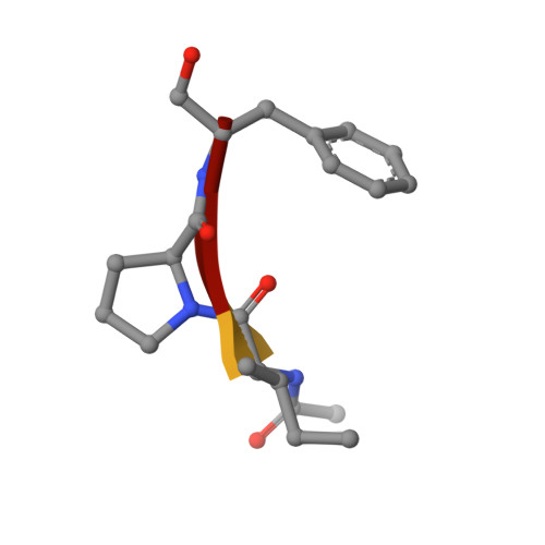
    Crystal structures of the serine-carboxyl proteinase from Pseudomonas sp. 101 (PSCP), complexed with a number of inhibitors, have been solved and refined at high- to atomic-level resolution. All of these inhibitors (tyrostatin, pseudo-tyrostatin, AcIPF, AcIAF, and chymostatin, as well as previously studied iodotyrostatin and pseudo-iodotyrostatin) make covalent bonds to the active site Ser287 through their aldehyde moieties, while their side chains occupy subsites S1-S4 of the enzyme. The mode of binding of the inhibitors is almost identical for their P1 and P2 side chains, while significant differences are observed for P3 and P4 (if present). Kinetic parameters for the binding of these nanomolar inhibitors to PSCP have been established and correlated with the observed mode of binding. The preferences of this enzyme for a larger side chain in P2 as well as Tyr or Phe in P1 are explained by the size, shape, and characteristics of the S2 and S1 regions of the protein structure, respectively. Networks of hydrogen bonds involving glutamic and aspartic acids have been analyzed for the atomic-resolution structure of the native enzyme. PSCP contains a calcium-binding site that consists of Asp328, Asp348, three amide carbonyl groups, and a water molecule, in almost perfect octahedral coordination. The presence of Ca(2+) cation is necessary for the activity of the enzyme.


  • Organizational Affiliation
    • Protein Structure Section, Macromolecular Crystallography Laboratory, and Intramural Research Support Program, SAIC Frederick, National Cancer Institute at Frederick, Frederick, Maryland 21702, USA. wlodawer@ncifcrf.gov

Macromolecule Content 

  • Total Structure Weight: 38.92 kDa 
  • Atom Count: 3,253 
  • Modeled Residue Count: 373 
  • Deposited Residue Count: 373 
  • Unique protein chains: 2

Macromolecules

Find similar proteins by:|  3D Structure
Entity ID: 1
MoleculeChains  Sequence LengthOrganismDetailsImage
SEDOLISIN369Pseudomonas sp. 101Mutation(s): 0 
Gene Names: pcp
EC: 3.4.21.100
UniProt
Find proteins for P42790 (Pseudomonas sp. (strain 101))
Explore P42790 
Go to UniProtKB:  P42790
Entity Groups
Sequence Clusters30% Identity50% Identity70% Identity90% Identity95% Identity100% Identity
UniProt GroupP42790
Sequence Annotations
Expand
Reference Sequence
Find similar proteins by:  Sequence   |   3D Structure  
Entity ID: 2
MoleculeChains  Sequence LengthOrganismDetailsImage
AIPF PEPTIDE INHIBITOR4synthetic constructMutation(s): 0 

Biologically Interesting Molecules (External Reference) 

1 Unique

Experimental Data & Validation

Experimental Data

  • Method: X-RAY DIFFRACTION
  • Resolution: 1.10 Å
  • R-Value Free:  0.157 (Depositor), 0.120 (DCC) 
  • R-Value Work:  0.132 (Depositor) 
  • R-Value Observed: 0.132 (Depositor) 
Space Group: P 62
Unit Cell:
Length ( Å )Angle ( ˚ )
a = 97.29α = 90
b = 97.29β = 90
c = 83.46γ = 120
Software Package:
Software NamePurpose
SCALEPACKdata scaling
SHELXLrefinement
PDB_EXTRACTdata extraction
HKL-2000data reduction

Structure Validation

View Full Validation Report


Entry History 

& Funding Information

Deposition Data


Funding OrganizationLocationGrant Number
Ministry of Education, Culture, Sports, Science and Technology (Japan)Japan13460043
National Institutes of Health/National Institute of Diabetes and Digestive and Kidney Disease (NIH/NIDDK)United StatesDK18865 & AI28571
National Institutes of Health/National Cancer Institute (NIH/NCI)United Statesintramural funds

Revision History  (Full details and data files)

  • Version 1.0: 2018-10-24
    Type: Initial release
  • Version 1.1: 2019-12-04
    Changes: Author supporting evidence
  • Version 1.2: 2023-10-11
    Changes: Data collection, Database references, Derived calculations, Refinement description