6M7Y | pdb_00006m7y

Dehydratase, NisB, bound to a non-eliminable substrate analog


Experimental Data Snapshot

  • Method: X-RAY DIFFRACTION
  • Resolution: 2.79 Å
  • R-Value Free: 
    0.270 (Depositor), 0.274 (DCC) 
  • R-Value Work: 
    0.185 (Depositor), 0.192 (DCC) 
  • R-Value Observed: 
    0.189 (Depositor) 

Starting Model: experimental
View more details

wwPDB Validation 3D Report Full Report

Validation slider image for 6M7Y

This is version 2.1 of the entry. See complete history

Literature

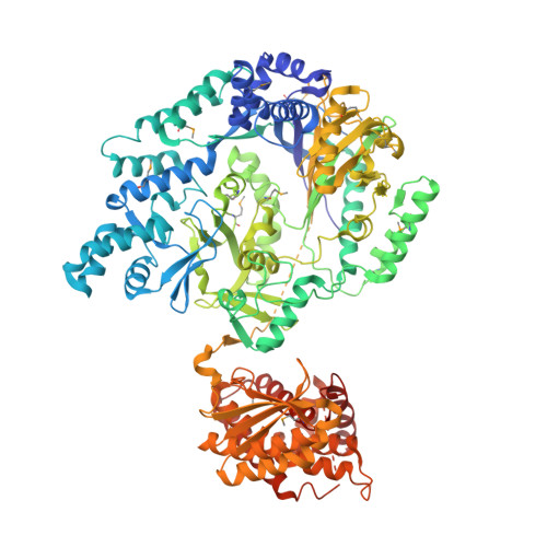
Characterization of glutamyl-tRNA-dependent dehydratases using nonreactive substrate mimics.

Bothwell, I.R.Cogan, D.P.Kim, T.Reinhardt, C.J.van der Donk, W.A.Nair, S.K.

(2019) Proc Natl Acad Sci U S A 116: 17245-17250

  • DOI: https://doi.org/10.1073/pnas.1905240116
  • Primary Citation Related Structures: 
    6EC7, 6EC8, 6M7Y

  • PubMed Abstract: 

    The peptide natural product nisin has been used as a food preservative for 6 decades with minimal development of resistance. Nisin contains the unusual amino acids dehydroalanine and dehydrobutyrine, which are posttranslationally installed by class I lanthipeptide dehydratases (LanBs) on a linear peptide substrate through an unusual glutamyl-tRNA-dependent dehydration of Ser and Thr. To date, little is known about how LanBs catalyze the transfer of glutamate from charged tRNA Glu to the peptide substrate, or how they carry out the subsequent elimination of the peptide-glutamyl adducts to afford dehydro amino acids. Here, we describe the synthesis of inert analogs that mimic substrate glutamyl-tRNA Glu and the glutamylated peptide intermediate, and determine the crystal structures of 2 LanBs in complex with each of these compounds. Mutational studies were used to characterize the function of the glutamylation and glutamate elimination active-site residues identified through the structural analysis. These combined studies provide insights into the mechanisms of substrate recognition, glutamylation, and glutamate elimination by LanBs to effect a net dehydration reaction of Ser and Thr.


  • Organizational Affiliation
    • Department of Chemistry, University of Illinois at Urbana-Champaign, Urbana, IL 61801.

Macromolecule Content 

  • Total Structure Weight: 244.58 kDa 
  • Atom Count: 16,392 
  • Modeled Residue Count: 1,932 
  • Deposited Residue Count: 2,060 
  • Unique protein chains: 2

Macromolecules

Find similar proteins by:|  3D Structure
Entity ID: 1
MoleculeChains  Sequence LengthOrganismDetailsImage
Nisin biosynthesis protein NisB
A, B
996Lactococcus lactis subsp. lactisMutation(s): 1 
Gene Names: nisB
UniProt
Find proteins for P20103 (Lactococcus lactis subsp. lactis)
Explore P20103 
Go to UniProtKB:  P20103
Entity Groups
Sequence Clusters30% Identity50% Identity70% Identity90% Identity95% Identity100% Identity
UniProt GroupP20103
Sequence Annotations
Expand
Reference Sequence
Find similar proteins by:|  3D Structure
Entity ID: 2
MoleculeChains  Sequence LengthOrganismDetailsImage
Lantibiotic
C, D
34Lactococcus lactisMutation(s): 4 
UniProt
Find proteins for P29559 (Lactococcus lactis subsp. lactis)
Explore P29559 
Go to UniProtKB:  P29559
Entity Groups
Sequence Clusters30% Identity50% Identity70% Identity90% Identity95% Identity100% Identity
UniProt GroupP29559
Sequence Annotations
Expand
Reference Sequence

Small Molecules

Modified Residues  1 Unique
IDChains TypeFormula2D DiagramParent
MSE
Query on MSE
A, B
L-PEPTIDE LINKINGC5 H11 N O2 SeMET
J9A
Query on J9A
C, D
L-PEPTIDE LINKINGC8 H15 N3 O5

--

Experimental Data & Validation

Experimental Data

  • Method: X-RAY DIFFRACTION
  • Resolution: 2.79 Å
  • R-Value Free:  0.270 (Depositor), 0.274 (DCC) 
  • R-Value Work:  0.185 (Depositor), 0.192 (DCC) 
  • R-Value Observed: 0.189 (Depositor) 
Space Group: P 1 21 1
Unit Cell:
Length ( Å )Angle ( ˚ )
a = 99.644α = 90
b = 107.143β = 109.57
c = 135.438γ = 90
Software Package:
Software NamePurpose
PHENIXrefinement
HKL-2000data reduction
HKL-2000data scaling
PHASERphasing

Structure Validation

View Full Validation Report


Entry History 

Deposition Data

Revision History  (Full details and data files)

  • Version 1.0: 2019-08-14
    Type: Initial release
  • Version 1.1: 2019-08-28
    Changes: Data collection, Database references
  • Version 1.2: 2019-09-04
    Changes: Data collection, Database references
  • Version 1.3: 2023-10-11
    Changes: Data collection, Database references, Refinement description
  • Version 2.0: 2023-11-15
    Changes: Atomic model, Data collection, Derived calculations
  • Version 2.1: 2024-11-13
    Changes: Structure summary