6M5B | pdb_00006m5b

X-ray crystal structure of cyclic-PIP and DNA complex in a reverse binding orientation


Experimental Data Snapshot

  • Method: X-RAY DIFFRACTION
  • Resolution: 1.50 Å
  • R-Value Free: 
    0.189 (Depositor), 0.212 (DCC) 
  • R-Value Work: 
    0.184 (Depositor), 0.196 (DCC) 
  • R-Value Observed: 
    0.184 (Depositor) 

wwPDB Validation 3D Report Full Report

Validation slider image for 6M5B

Ligand Structure Quality Assessment 


This is version 1.1 of the entry. See complete history

Literature

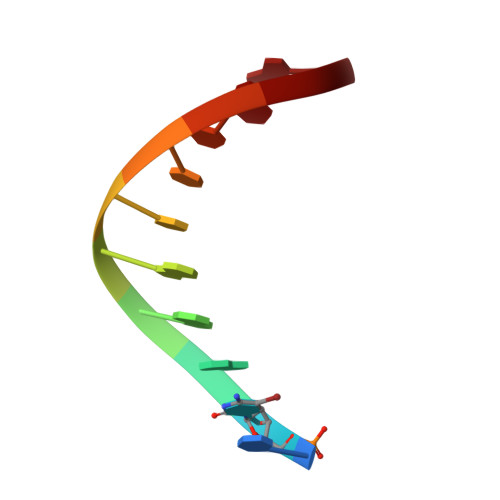
X-ray Crystal Structure of a Cyclic-PIP-DNA Complex in the Reverse-Binding Orientation.

Abe, K.Hirose, Y.Eki, H.Takeda, K.Bando, T.Endo, M.Sugiyama, H.

(2020) J Am Chem Soc 142: 10544-10549

  • DOI: https://doi.org/10.1021/jacs.0c03972
  • Primary Citation Related Structures: 
    6M5B

  • PubMed Abstract: 

    Elucidation of the details of the associating mode is one of the major concerns for the precise design of DNA-binding molecules that are used for gene regulation. Pyrrole-imidazole polyamide (PIP) is a well-established synthetic DNA-binding molecule that has sequence-specificity for duplex DNA. By the design of the sequence of pyrrole, imidazole, and other synthetic units, PIP is bound to the target DNA sequence selectively. Here, we report the X-ray crystal structure of newly synthesized chiral cyclic PIP (cPIP) complexed with DNA at 1.5 Å resolution and reveal that cPIP binds in the reverse orientation in the DNA minor groove. Analysis of the crystal structure revealed that the positions of the hydrogen bonds between the bases and the pyrrole-imidazole moieties of cPIP were similar for both forward- and reverse-binding orientations and that the distortion of the B-form DNA structure caused by cPIP binding was also similar for both orientations. We further found that new hydrogen bonds formed between the amino groups on the γ-turn units and DNA at both ends of the cPIP molecule. Additionally, by comparing the reverse PIP orientation with the forward orientation, we could clarify that the cause of the preference toward the reverse orientation in the S -form cPIP as used in this study is the overall conformation of the cPIP-DNA complex, particularly the configuration of hydrogen bonds. These results thus provide an explanation for the different stereoselectivity of cPIP binding in the minor groove.


  • Organizational Affiliation
    • Department of Chemistry, Graduate School of Science, Kyoto University, Kitashirakawa-oiwakecho, Sakyo-ku, Kyoto 606-8502, Japan.

Macromolecule Content 

  • Total Structure Weight: 3.83 kDa 
  • Atom Count: 298 
  • Modeled Residue Count: 10 
  • Deposited Residue Count: 10 
  • Unique nucleic acid chains: 1

Macromolecules

Find similar nucleic acids by:  Sequence
Entity ID: 1
MoleculeChains LengthOrganismImage
DNA (5'-D(*CP*(CBR)P*AP*GP*GP*CP*CP*TP*GP*G)-3')10synthetic construct
Sequence Annotations
Expand
Reference Sequence

Small Molecules

Ligands 4 Unique
IDChains Name / Formula / InChI Key2D Diagram3D Interactions
S7E
(Subject of Investigation/LOI)

Query on S7E


Download:Ideal Coordinates CCD File
B [auth A]4-[[4-[(4-azanyl-1-methyl-pyrrol-2-yl)carbonylamino]-1-methyl-pyrrol-2-yl]carbonylamino]-~{N}-[2-[[(3~{S})-3-azanyl-4-oxidanylidene-butyl]carbamoyl]-1-methyl-imidazol-4-yl]-1-methyl-imidazole-2-carboxamide
C26 H32 N12 O5
UZFBAFZKKPMVCV-AWEZNQCLSA-N
EDO

Query on EDO


Download:Ideal Coordinates CCD File
E [auth A]1,2-ETHANEDIOL
C2 H6 O2
LYCAIKOWRPUZTN-UHFFFAOYSA-N
MG

Query on MG


Download:Ideal Coordinates CCD File
C [auth A]MAGNESIUM ION
Mg
JLVVSXFLKOJNIY-UHFFFAOYSA-N
NA

Query on NA


Download:Ideal Coordinates CCD File
D [auth A]SODIUM ION
Na
FKNQFGJONOIPTF-UHFFFAOYSA-N

Experimental Data & Validation

Experimental Data

  • Method: X-RAY DIFFRACTION
  • Resolution: 1.50 Å
  • R-Value Free:  0.189 (Depositor), 0.212 (DCC) 
  • R-Value Work:  0.184 (Depositor), 0.196 (DCC) 
  • R-Value Observed: 0.184 (Depositor) 
Space Group: I 41 3 2
Unit Cell:
Length ( Å )Angle ( ˚ )
a = 82.235α = 90
b = 82.235β = 90
c = 82.235γ = 90
Software Package:
Software NamePurpose
PHENIXrefinement
XDSdata reduction
XDSdata scaling
SOLVEphasing

Structure Validation

View Full Validation Report


Ligand Structure Quality Assessment 


Entry History 

& Funding Information

Deposition Data


Funding OrganizationLocationGrant Number
Ministry of Education, Culture, Sports, Science and Technology (Japan)Japan18KK0139
Ministry of Education, Culture, Sports, Science and Technology (Japan)Japan16H06356

Revision History  (Full details and data files)

  • Version 1.0: 2020-06-24
    Type: Initial release
  • Version 1.1: 2024-03-27
    Changes: Data collection, Database references, Derived calculations