6M4Y | pdb_00006m4y

Structure of a R371A mutant of a Group II PLP dependent decarboxylase from Methanocaldococcus jannaschii


Experimental Data Snapshot

  • Method: X-RAY DIFFRACTION
  • Resolution: 2.10 Å
  • R-Value Free: 
    0.200 (Depositor), 0.200 (DCC) 
  • R-Value Work: 
    0.164 (Depositor), 0.165 (DCC) 
  • R-Value Observed: 
    0.165 (Depositor) 

Starting Model: experimental
View more details

wwPDB Validation 3D Report Full Report

Validation slider image for 6M4Y

This is version 1.2 of the entry. See complete history

Literature

Crystallographic Snapshots of the Dunathan and Quinonoid Intermediates provide Insights into the Reaction Mechanism of Group II Decarboxylases.

Gayathri, S.C.Manoj, N.

(2020) J Mol Biology 432: 166692-166692

  • DOI: https://doi.org/10.1016/j.jmb.2020.10.026
  • Primary Citation Related Structures: 
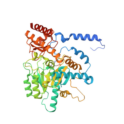
    6M4Y

  • PubMed Abstract: 

    PLP-dependent enzymes catalyze a plethora of chemical reactions affecting diverse physiological functions. Here we report the structural determinants of the reaction mechanism in a Group II PLP-dependent decarboxylase by assigning two early intermediates. The in-crystallo complexes of the PLP bound form, and the Dunathan and quinonoid intermediates, allowed direct observation of the active site interactions. The structures reveal that a subtle rearrangement of a conserved Arg residue in concert with a water-mediated interaction with the carboxylate of the Dunathan intermediate, appears to directly stabilize the alignment and facilitate the release of CO 2 to yield the quinonoid. Modeling indicates that the conformational change of a dynamic catalytic loop to a closed form controls a conserved network of hydrogen bond interactions between catalytic residues to protonate the quinonoid. Our results provide a structural framework to elucidate mechanistic roles of residues that govern reaction specificity and catalysis in PLP-dependent decarboxylation.


  • Organizational Affiliation
    • Department of Biotechnology, Bhupat and Jyoti Mehta School of Biosciences, Indian Institute of Technology Madras, Chennai 600036, India.

Macromolecule Content 

  • Total Structure Weight: 48.14 kDa 
  • Atom Count: 3,344 
  • Modeled Residue Count: 389 
  • Deposited Residue Count: 415 
  • Unique protein chains: 1

Macromolecules

Find similar proteins by:|  3D Structure
Entity ID: 1
MoleculeChains  Sequence LengthOrganismDetailsImage
L-tyrosine/L-aspartate decarboxylase415Methanocaldococcus jannaschii DSM 2661Mutation(s): 1 
Gene Names: mfnAMJ0050
EC: 4.1.1.11 (PDB Primary Data), 4.1.1.25 (PDB Primary Data)
UniProt
Find proteins for Q60358 (Methanocaldococcus jannaschii (strain ATCC 43067 / DSM 2661 / JAL-1 / JCM 10045 / NBRC 100440))
Explore Q60358 
Go to UniProtKB:  Q60358
Entity Groups
Sequence Clusters30% Identity50% Identity70% Identity90% Identity95% Identity100% Identity
UniProt GroupQ60358
Sequence Annotations
Expand
Reference Sequence

Experimental Data & Validation

Experimental Data

  • Method: X-RAY DIFFRACTION
  • Resolution: 2.10 Å
  • R-Value Free:  0.200 (Depositor), 0.200 (DCC) 
  • R-Value Work:  0.164 (Depositor), 0.165 (DCC) 
  • R-Value Observed: 0.165 (Depositor) 
Space Group: P 43 21 2
Unit Cell:
Length ( Å )Angle ( ˚ )
a = 98.44α = 90
b = 98.44β = 90
c = 121.7γ = 90
Software Package:
Software NamePurpose
Aimlessdata scaling
PHENIXrefinement
PDB_EXTRACTdata extraction
iMOSFLMdata reduction
PHENIXphasing

Structure Validation

View Full Validation Report


Entry History 

Deposition Data

Revision History  (Full details and data files)

  • Version 1.0: 2020-12-02
    Type: Initial release
  • Version 1.1: 2020-12-16
    Changes: Database references
  • Version 1.2: 2023-11-29
    Changes: Data collection, Database references, Refinement description