6M3L | pdb_00006m3l

Crystal structure of the R.PabI(Y68F-K154A)-dsDNA(nonspecific) complex


Experimental Data Snapshot

  • Method: X-RAY DIFFRACTION
  • Resolution: 2.75 Å
  • R-Value Free: 
    0.306 (Depositor), 0.309 (DCC) 
  • R-Value Work: 
    0.283 (Depositor), 0.288 (DCC) 
  • R-Value Observed: 
    0.284 (Depositor) 

Starting Model: experimental
View more details

wwPDB Validation 3D Report Full Report

Validation slider image for 6M3L

This is version 1.4 of the entry. See complete history

Literature

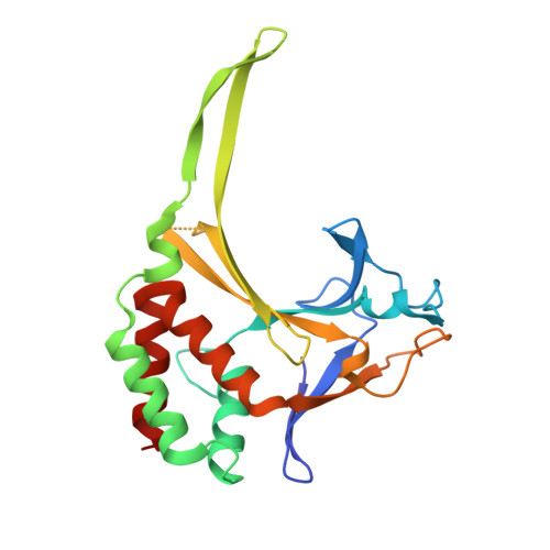
Distortion of double-stranded DNA structure by the binding of the restriction DNA glycosylase R.PabI.

Miyazono, K.I.Wang, D.Ito, T.Tanokura, M.

(2020) Nucleic Acids Res 48: 5106-5118

  • DOI: https://doi.org/10.1093/nar/gkaa184
  • Primary Citation Related Structures: 
    6L2N, 6L2O, 6M3L

  • PubMed Abstract: 

    R.PabI is a restriction DNA glycosylase that recognizes the sequence 5'-GTAC-3' and hydrolyses the N-glycosidic bond of adenine in the recognition sequence. R.PabI drastically bends and unwinds the recognition sequence of double-stranded DNA (dsDNA) and flips the adenine and guanine bases in the recognition sequence into the catalytic and recognition sites on the protein surface. In this study, we determined the crystal structure of the R.PabI-dsDNA complex in which the dsDNA is drastically bent by the binding of R.PabI but the base pairs are not unwound. This structure is predicted to be important for the indirect readout of the recognition sequence by R.PabI. In the complex structure, wedge loops of the R.PabI dimer are inserted into the minor groove of dsDNA to stabilize the deformed dsDNA structure. A base stacking is distorted between the two wedge-inserted regions. R.PabI is predicted to utilize the distorted base stacking for the detection of the recognition sequence.


  • Organizational Affiliation
    • Department of Applied Biological Chemistry, Graduate School of Agricultural and Life Sciences, The University of Tokyo, 1-1-1 Yayoi Bunkyo-ku, Tokyo 113-8657, Japan.

Macromolecule Content 

  • Total Structure Weight: 57.71 kDa 
  • Atom Count: 3,342 
  • Modeled Residue Count: 390 
  • Deposited Residue Count: 463 
  • Unique protein chains: 1
  • Unique nucleic acid chains: 1

Macromolecules


Find similar proteins by:|  3D Structure
Entity ID: 1
MoleculeChains  Sequence LengthOrganismDetailsImage
RE_R_Pab1 domain-containing protein
A, B
220Pyrococcus abyssi GE5Mutation(s): 2 
Gene Names: PAB0105
UniProt
Find proteins for G8ZFZ3 (Pyrococcus abyssi (strain GE5 / Orsay))
Explore G8ZFZ3 
Go to UniProtKB:  G8ZFZ3
Entity Groups
Sequence Clusters30% Identity50% Identity70% Identity90% Identity95% Identity100% Identity
UniProt GroupG8ZFZ3
Sequence Annotations
Expand
Reference Sequence
Find similar nucleic acids by:  Sequence
Entity ID: 2
MoleculeChains LengthOrganismImage
DNA (5'-D(*CP*GP*CP*AP*TP*CP*GP*AP*TP*TP*CP*AP*GP*AP*AP*TP*CP*GP*AP*TP*GP*CP*G)-3')23synthetic construct
Sequence Annotations
Expand
Reference Sequence

Experimental Data & Validation

Experimental Data

  • Method: X-RAY DIFFRACTION
  • Resolution: 2.75 Å
  • R-Value Free:  0.306 (Depositor), 0.309 (DCC) 
  • R-Value Work:  0.283 (Depositor), 0.288 (DCC) 
  • R-Value Observed: 0.284 (Depositor) 
Space Group: P 32 2 1
Unit Cell:
Length ( Å )Angle ( ˚ )
a = 84.153α = 90
b = 84.153β = 90
c = 140.53γ = 120
Software Package:
Software NamePurpose
PHENIXrefinement
PDB_EXTRACTdata extraction
XDSdata reduction
XDSdata scaling
MOLREPphasing

Structure Validation

View Full Validation Report


Entry History 

Deposition Data

Revision History  (Full details and data files)

  • Version 1.0: 2020-03-18
    Type: Initial release
  • Version 1.1: 2020-04-01
    Changes: Derived calculations
  • Version 1.2: 2020-04-15
    Changes: Database references
  • Version 1.3: 2020-05-27
    Changes: Database references
  • Version 1.4: 2023-11-29
    Changes: Data collection, Database references, Refinement description